
Page 312 - Read Online
P. 312
Mok et al. Mini-invasive Surg 2019;3:40 I http://dx.doi.org/10.20517/2574-1225.2019.39 Page 3 of 9
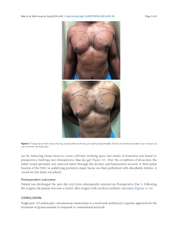
Figure 1. Preoperative front views showing preoperative markings and right gynaecomastia. Extent of planned dissection was marked out
(as shown by red markings)
out by retracting breast tissue to create sufficient working space and extent of dissection was based on
preoperative markings and intraoperative blue dye gel [Figure 10]. After the completion of dissection, the
entire breast specimen was removed intact through the incision and haemostasis secured. A three-point
fixation of the NAC to underlying pectoralis major fascia was then performed with absorbable stitches. A
closed suction drain was placed.
Postoperative outcomes
Patient was discharged the next day and drain subsequently removed on Postoperative Day 5. Following
the surgery, the patient was seen a month after surgery with excellent aesthetic outcomes [Figures 11-13].
CONCLUSION
Single-port 3D endoscopic subcutaneous mastectomy is a novel and aesthetically superior approach for the
treatment of gynaecomastia if compared to conventional methods.