
Page 313 - Read Online
P. 313
Page 4 of 9 Mok et al. Mini-invasive Surg 2019;3:40 I http://dx.doi.org/10.20517/2574-1225.2019.39
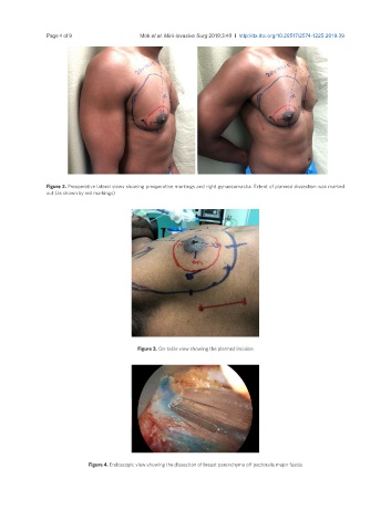
Figure 2. Preoperative lateral views showing preoperative markings and right gynaecomastia. Extent of planned dissection was marked
out (as shown by red markings)
Figure 3. On-table view showing the planned incision
Figure 4. Endoscopic view showing the dissection of breast parenchyma off pectoralis major fascia