
Page 31 - Read Online
P. 31
Ewbank et al. Mini-invasive Surg 2018;2:5 I http://dx.doi.org/10.20517/2574-1225.2017.51 Page 3 of 5
A B C
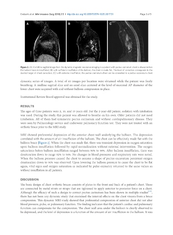
Figure 2. (A) A midline sagittal image from the dynamic magnetic resonance imaging in a patient with pectus carinatum chest is shown before
the balloon brace is insufflated; (B) with sufficient insufflation of the balloon, the chest is made flat. This level of correction corresponds to the
desired target of chest correction; (C) with extreme insufflation, the pectus carinatum chest can be converted to a pectus excavatum chest
dynamic series of images. A total of 60 images per location were obtained while the patient was freely
breathing. A midline sagittal slice and an axial slice centered at the level of maximal AP diameter of the
lower chest were acquired with and without balloon compression in place.
Institutional Review Board approval was obtained for the study.
RESULTS
The ages of three patients were 2, 10, and 15 years old. For the 2-year-old patient, sedation with intubation
was used. During the study, this patient was allowed to breathe on his own. Older patients did not need
intubation. All of them had symmetric pectus carinatum and without cardiopulmonary disease. They
were seen by Pulmonology service and underwent pulmonary function test. They were not treated with an
orthotic brace prior to the MRI study.
MRI showed preferential depression of the anterior chest wall underlying the balloon. The depression
correlated with the amount of air insufflation of the balloon. The chest can be effectively made flat with the
balloon brace [Figure 2]. When the chest was made flat, there was transient depression in oxygen saturation
upon balloon insufflation followed by rapid normalization without external intervention. The oxygen
saturations before balloon insufflation ranged between 96% to 98%. After balloon insufflation, there was
desaturation down to range 92% to 94%. No changes in blood pressures and respiratory rate were noted.
When the balloon pressure caused the chest to assume a shape of pectus excavatum persistent oxygen
desaturation down to 90% was observed. Upon lowering the balloon pressure to cause the chest to be flat
again, vital signs and oxygen saturation as indicated by pulse oximetry returned to the same values as
without insufflation in all patients.
DISCUSSION
The basic design of chest orthotic braces consists of plates in the front and back of a patient’s chest. These
are connected by metal struts or straps that are tightened to apply anterior to posterior force on a chest.
[3-5]
Although the efficacy of such a design to correct pectus carinatum has been shown in multiple studies ,
there has not been any dynamic study that examined the internal effects on the chest viscera from a brace
compression. This dynamic MRI study showed that preferential compression of anterior chest did not alter
blood pressure, pulse, or pulmonary function. The finding indicates that the patient’s cardiac and pulmonary
function can compensate for the compression. The chest wall area under the balloon is clearly observed to
be depressed, and the level of depression is a function of the amount of air insufflation in the balloon. It was