
Page 30 - Read Online
P. 30
Page 2 of 5 Ewbank et al. Mini-invasive Surg 2018;2:5 I http://dx.doi.org/10.20517/2574-1225.2017.51
A B C
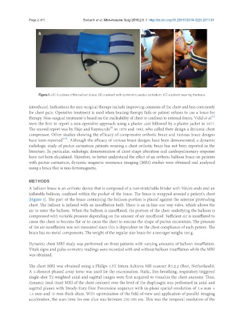
Figure 1. (A) A picture of the balloon brace; (B) a patient with symmetric pectus carinatum; (C) a patient wearing the brace
introduced. Indications for non-surgical therapy include improving cosmesis of the chest and less commonly
for chest pain. Operative treatment is used when bracing therapy fails or patient refuses to use a brace for
[1]
therapy. Non-surgical treatment is based on the malleability of chest to conform to external forces. Vidal et al.
were the first to report a non-operative approach using a plaster cast followed by a plaster jacket in 1977.
[2]
The second report was by Haje and Raymundo in 1979 and 1992, who called their design a dynamic chest
compressor. Other studies showing the efficacy of compressive orthotic brace and various brace designs
[3-5]
have been reported . Although the efficacy of various brace designs have been demonstrated, a dynamic
radiologic study of pectus carinatum patients wearing a chest orthotic brace has not been reported in the
literature. In particular, radiologic demonstration of chest shape alteration and cardiopulmonary response
have not been elucidated. Therefore, to better understand the effect of an orthotic balloon brace on patients
with pectus carinatum, dynamic magnetic resonance imaging (MRI) studies were obtained and analyzed
using a brace that is non-ferromagnetic.
METHODS
A balloon brace is an orthotic device that is composed of a non-stretchable binder with Velcro ends and an
inflatable balloon, confined within the pocket of the brace. The brace is wrapped around a patient’s chest
[Figure 1]. The part of the brace containing the balloon portion is placed against the anterior protruding
chest. The balloon is inflated with an insufflation bulb. There is an in-line one way valve, which allows the
air to enter the balloon. When the balloon is insufflated, the portion of the chest underlying the balloon is
compressed with variable pressure depending on the amount of air insufflated. Sufficient air is insufflated to
cause the chest to become flat or to cause the chest to assume the shape of pectus excavatum. The pressure
of the air insufflation was not measured since this is dependent on the chest compliance of each patient. The
brace has no metal components. The weight of the regular size brace for a teenager weighs 150 g.
Dynamic chest MRI study was performed on three patients with varying amounts of balloon insufflation.
Vitals signs and pulse oximetry readings were recorded with and without balloon insufflation while the MRI
was obtained.
The chest MRI was obtained using a Philips 1.5T Intera-Achieva MR scanner R3.2.2 (Best, Netherlands).
A 4-element phased-array torso was used for the examination. Static, free-breathing, respiratory-triggered
single-shot T2-weighted axial and sagittal images were first acquired to visualize the chest anatomy. Then,
dynamic (real-time) MRI of the chest centered over the level of the diaphragm was performed in axial and
sagittal planes with Steady-State Free Precession sequence with in-plane spatial resolution of 1.4 mm ×
1.4 mm and 10 mm thick slices. With optimization of the field-of-view and application of parallel imaging
acceleration, the scan time for one slice was between 220-290 ms. This was the temporal resolution of the