
Page 36 - Read Online
P. 36
Priego et al. Mini-invasive Surg 2018;2:6 I http://dx.doi.org/10.20517/2574-1225.2018.01 Page 3 of 7
A B
Figure 1. Gastrointestinal stromal tumor near the esophagogastric junction observed in oral endoscopy (A) and computerized tomography scan (B)
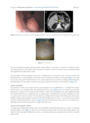
Figure 2. Trocar placement
First, we started the procedure after inserting a supraumbilical 10-mm port. A second 10-mm ballon trocar
was placed along the left midclavicular line and a third port, sized 5-mm ballon trocar was placed along
the epigastric line, under direct vision.
The procedure started with the incision of a suitable point on the gastric wall, which served for the
introduction of a 12-mm balloon trocar. This step was performed using the Ultracision [Figure 3A]. This
trocar was used to allow the introduction of a 10-mm scope and also to allow the sealing of the stomach to
the abdominal wall. The other 10 and 5 mm balloon trocars were inserted into the stomach [Figure 3B].
Tumor resection
Once balloon trocars were inside stomach, pneumogastrum was established (4-6 mmHg).The location
of the tumor was confirmed after the introduction of the scope [Figure 4A]. For tumors near the EGJ,
precaution was taken not to involve the EGJ itself. For that, the EGJ needed to be clearly identified either
by insertion of the tip of a nasogastric tube or a gastroscope. To facilitate the resection, sometimes we used
a tractive suture into the tumor. The resection was performed by means of the Ultracision (Harmonic
Scalpel; Ethicon Endo-Surgery, Cincinnati, OH, USA), making sure to leave a 1-cm of clear tissue around
the lesion without breach of the capsule [Figure 4B]. Once the tumor has been removed, we introduced an
Endobag, and retrieved the specimen through the ballon trocar.
Closure of the gastric defect
A single layer of interrupted sutures with non-absorbable material (Ethibond 2/0) was used to closure the
posterior gastric wall [Figure 5]. The trocar was then be retrieved under vision and the anterior gastric wall
sutured with interrupted Ethibond 2/0 suture. A methylene blue test was performed in order to exclude
gastric leaks.