
Page 125 - Read Online
P. 125
Dantas Idiopathic and chagasic achalasia
body . Using high resolution manometry achalasia esophagus, including absent or partial relaxation of the
[3]
patients may be classified as type I, when there are no lower esophageal sphincter, esophageal aperistlsis,
contractions in esophageal body during swallows, type and megaesophagus the loss of esophageal intrinsic
II, characterized by pan-esophageal pressurization, or innervations may not be the same [11-13] .
type III when there are high-amplitude simultaneous
contractions in the distal esophagus . While in idiopathic achalasia neural destruction has
[3]
been suggested to be more intense in inhibitory
The etiology of achalasia is unknown in most cases nerves than in excitatory nerves, in achalasia caused
around the world, and may be multifactorial, including by Trypanosoma cruzi infection neural impairment
autoimmune, genetic and viral factors . In idiopathic involves both inhibitory and excitatory innervations.
[2]
achalasia, there are evidences of autoimmune, genetic Consequently lower esophageal sphincter pressure is
and viral etiology, due to the presence of specific frequently increased in idiopathic achalasia [14-17] and
autoantibodies associated with neuronal damage, frequently decreased in Chagas’ disease [16-19] which
occasional incidence in members of the same family, may explain the variation in the lower esophageal
and presence of previous viral infection in these sphincter pressure [20] , and the heterogeneity seen in
patients . The disease occurs with an annual incidence these patients [21] .
[2]
of 1 in 100,000 and a prevalence of 10 in 100,000 .
[4]
Previous studies have reported differences in esophageal
Achalasia may be caused by infection by the response to gastrin [14,15,18] and to atropine [15,22] . These
hemoflagellate protozoan Trypanosoma cruzi [5,6] mechanisms have not been completely elucidated
which affects millions of people in Latin America and in Chagas’ disease [11,13] [Table 1]. Although studies
has been increasingly reported in the United States on idiopathic achalasia have demonstrated a partial
[7]
and Europe . This parasitic infection is the cause of opening of the upper esophageal sphincter with
[8]
Chagas’ disease, and is characterized by myenteric increased residual pressure during swallow [23] , these
inflammation, absent myenteric ganglion cells and features have not been fully demonstrated in Chagas’
myenteric neural fibrosis. These lesions are restricted disease [12,13] . The time between pharyngeal contraction
to the esophagus in idiophatic achalasia , and may and proximal esophageal contraction (5 cm distance)
[2]
be seen in all digestive tract in Chagas’ disease [5,6,9,10] . after wet swallows in patients with megaesophagus
In Latin America Chagas’ disease has an incidence is increased in Chagas’ disease but not in idiopathic
from 1,000 in 100,000 to 4,000 in 100,000, however achalasia [24] . Contractions in the esophageal body are
the number is decreasing, as 18 million in 1991 to 5.7 not of the same intensity, and tend to be more intense
million in 2010 . It is estimated that 300,000 infected in patients with idiopathic achalasia [19,25] . In addition
[9]
immigrants are living in United States . From 7% to epiphrenic diverticula is more frequent on idiopathic
[9]
10% of the infected individuals will have achalasia . achalasia (3.6% to 7.4%) than in Chagas’ disease
[5]
(1.5%) [13] . Also, high prevalence of circulating antibodies
DIFFERENCES BETWEEN IDIOPATHIC AND against M2 acethilcholine muscarinic receptor has
CHAGAS’ DISEASE ACHALASIA been found in Chagas’ disease patients with achalasia
(84%), compared with patients with idiopathic achalasia
Although both diseases cause the same alteration in the (28%) [26] .
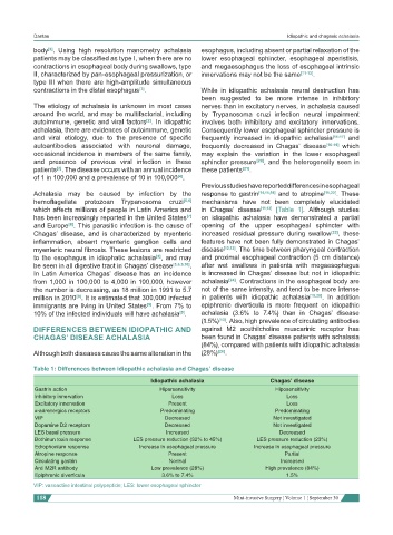
Table 1: Differences between idiopathic achalasia and Chagas’ disease
Idiopathic achalasia Chagas’ disease
Gastrin action Hipersensitivity Hiposensitivity
Inhibitory innervation Loss Loss
Excitatory innervation Present Loss
α-adrenergics receptors Predominating Predominating
VIP Decreased Not investigated
Dopamine D2 receptors Decreased Not investigated
LES basal pressure Increased Decreased
Bothinun toxin response LES pressure reduction (32% to 45%) LES pressure reduction (23%)
Edrophonium response Increase in esophageal pressure Increase in esophageal pressure
Atropine response Present Partial
Circulating gastrin Normal Increased
Anti M2R antibody Low prevalence (28%) High prevalence (84%)
Epiphrenic diverticula 3.6% to 7.4% 1.5%
VIP: vasoactive intestinal polypeptide; LES: lower esophageal sphincter
118 Mini-invasive Surgery ¦ Volume 1 ¦ September 30