
Page 12 - Read Online
P. 12
Page 6 of 11 Kikuchi et al. Mini-invasive Surg 2024;8:8 https://dx.doi.org/10.20517/2574-1225.2023.88
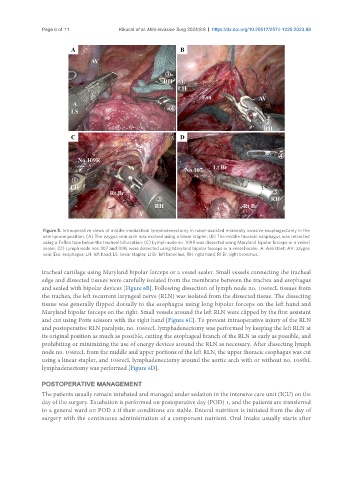
Figure 5. Intraoperative views of middle mediastinal lymphadenectomy in robot-assisted minimally invasive esophagectomy in the
semi-prone position. (A) The azygos vein arch was excised using a linear stapler; (B) The middle thoracic esophagus was retracted
using a Teflon tape below the tracheal bifurcation; (C) Lymph node no. 109R was dissected using Maryland bipolar forceps or a vessel
sealer; (D) Lymph node nos. 107 and 109L were dissected using Maryland bipolar forceps or a vessel sealer. A: Assistant; AV: azygos
vein; Eso: esophagus; LH: left hand; LS: linear stapler; Lt Br: left bronchus; RH: right hand; Rt Br: right bronchus.
tracheal cartilage using Maryland bipolar forceps or a vessel sealer. Small vessels connecting the tracheal
edge and dissected tissues were carefully isolated from the membrane between the trachea and esophagus
and sealed with bipolar devices [Figure 6B]. Following dissection of lymph node no. 106recL tissues from
the trachea, the left recurrent laryngeal nerve (RLN) was isolated from the dissected tissue. The dissecting
tissue was generally flipped dorsally to the esophagus using long bipolar forceps on the left hand and
Maryland bipolar forceps on the right. Small vessels around the left RLN were clipped by the first assistant
and cut using Potts scissors with the right hand [Figure 6C]. To prevent intraoperative injury of the RLN
and postoperative RLN paralysis, no. 106recL lymphadenectomy was performed by keeping the left RLN at
its original position as much as possible, cutting the esophageal branch of the RLN as early as possible, and
prohibiting or minimizing the use of energy devices around the RLN as necessary. After dissecting lymph
node no. 106recL from the middle and upper portions of the left RLN, the upper thoracic esophagus was cut
using a linear stapler, and 106recL lymphadenectomy around the aortic arch with or without no. 106tbL
lymphadenectomy was performed [Figure 6D].
POSTOPERATIVE MANAGEMENT
The patients usually remain intubated and managed under sedation in the intensive care unit (ICU) on the
day of the surgery. Extubation is performed on postoperative day (POD) 1, and the patients are transferred
to a general ward on POD 2 if their conditions are stable. Enteral nutrition is initiated from the day of
surgery with the continuous administration of a component nutrient. Oral intake usually starts after