
Page 76 - Read Online
P. 76
Page 8 of 11 Suwa et al. Mini-invasive Surg 2022;6:20 https://dx.doi.org/10.20517/2574-1225.2021.123
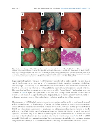
Figure 2. (A) An endoscopic white light image showing adenocarcinoma, 3rd portion, 0-IIa, cT1a (M), 12 mm; (B) endoscopic image
showing indigo carmine spraying; (C) filling the lumen with water after air deflation; (D) snaring with 15 mm snare; (E) mucosal defect
after UEMR where the procedure time was 3 min; (F) complete prophylactic clipping with two clips; (G) resected specimen shown as a
white light image; (H) resected specimen shown using narrow band imaging; and (I) histopathological image by hematoxylin and eosin
stain. The histopathological diagnosis was well differentiated tubular adenocarcinoma, pT1a (M), pHMX, pVM0.
Regarding the long-term outcomes, 52 of 76 lesions were followed up endoscopically for more than a
month. Local recurrence was observed in 3 of 52 lesions (4.8%) during a median observation period of two
months (range, 1-37 months). Of the three lesions with local recurrence, two lesions were treated with re-
UEMR and one lesion was followed up without additional treatment due to the patient’s general condition.
The procedural and long-term outcomes that were reported by Yamasaki et al. and our institution are
[19]
described in Table 3. A previous report and our data show that, although the R0 resection rate was low, the
recurrence rate was not as high (less than 5%). Importantly, the recurrent lesions were treatable by re-
endoscopic resection; however, this requires further evaluation with long-term follow-up data.
The advantages of UEMR include a relatively short procedure time and the ability to treat larger (> 10 mm)
and cancerous lesions. The disadvantages of UEMR are the low R0 resection rate, which is common to
lesions in both the colon and the duodenum. With the above results, we believe that the good indications of
UEMR are: (1) duodenal adenomas 10-20 mm in size; and (2) intramucosal carcinomas ≤ 20 mm in size. In
addition, a modified UEMR, partial submucosal injection UEMR (PI-UEMR), in which submucosal
injection is performed only on the difficult side (mostly anal side), has been reported to be effective in the
[26]
treatment of duodenal tumors (en bloc resection rate, 97%; R0 resection rate, 83%) . An RCT of UEMR
versus PI-UEMR with a primary endpoint of en bloc resection rate with pathologically confirmed negative
margins without conversion to ESD (R0 resection rate) is currently underway and the results are awaited.