
Page 74 - Read Online
P. 74
Page 6 of 11 Suwa et al. Mini-invasive Surg 2022;6:20 https://dx.doi.org/10.20517/2574-1225.2021.123
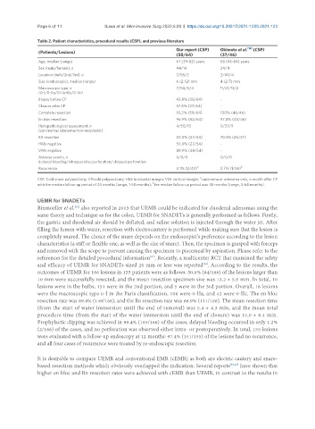
Table 2. Patient characteristics, procedural results (CSP), and previous literature
Our report (CSP) Okimoto et al. [12] (CSP)
(Patients/Lesions)
(58/64) (37/46)
Age, median (range) 67 (39-82) years 68 (46-84) years
Sex (male/female), n 44/14 24/11
Location (bulb/2nd/3rd), n 7/55/2 2/40/4
Size (endoscopic), median (range) 6 (2-12) mm 4 (2-7) mm
Macroscopic type, n 7/50/3/4 11/21/12/2
(0-I/0-IIa/0-IIa+IIc/0-IIc)
Biopsy before CP 43.8% (28/64) -
Closure after CP 57.8% (37/64) -
Complete resection 92.2% (59/64) 100% (46/46)
En bloc resection 96.9% (62/64) 97.8% (45/46)
Histopathological assessment, n 4/50/10 0/37/9
(carcinoma/adenoma/non-neoplastic)
R0 resection 50.0% (27/54) 70.3% (26/37)
HMs negative 50.0% (27/54) -
VMs negative 88.9% (48/54) -
Adverse events, n 0/0/0 0/0/0
delayed bleeding/intraoperative perforation/ delayed perforation
a b
Recurrence 4.1% (2/49) 2.7% (1/36)
a
CSP: Cold snare polypectomy; CP:cold polypectomy; HM: horizontal margin; VM: vertical margin; carcinoma or adenoma only, a month after CP
b
with the median follow-up period of 24 months (range, 1-58 months); the median follow-up period was 48 months (range, 3-64 months).
UEMR for SNADETs
Binmoeller et al. also reported in 2013 that UEMR could be indicated for duodenal adenomas using the
[20]
same theory and technique as for the colon. UEMR for SNADETs is generally performed as follows. Firstly,
the gastric and duodenal air should be deflated, and saline solution is injected through the water jet. After
filling the lumen with water, resection with electrocautery is performed while making sure that the lesion is
completely snared. The choice of the snare depends on the endoscopist’s preference according to the lesion
characteristics (a stiff or flexible one, as well as the size of snare). Then, the specimen is grasped with forceps
and removed with the scope to prevent causing the specimen to piecemeal by aspiration. Please refer to the
references for the detailed procedural information . Recently, a multicenter RCT that examined the safety
[21]
and efficacy of UEMR for SNADETs sized 20 mm or less was reported . According to the results, the
[22]
outcomes of UEMR for 166 lesions in 155 patients were as follows: 50.6% (84/166) of the lesions larger than
10 mm were successfully resected, and the mean resection specimen size was 13.2 ± 5.0 mm. In total, 10
lesions were in the bulbs, 151 were in the 2nd portion, and 5 were in the 3rd portion. Overall, 18 lesions
were the macroscopic type 0-I in the Paris classification, 106 were 0-IIa, and 42 were 0-IIc. The en bloc
resection rate was 89.8% (149/166), and the R0 resection rate was 66.9% (111/166). The mean resection time
(from the start of water immersion until the end of removal) was 5.4 ± 4.3 min, and the mean total
procedure time (from the start of the water immersion until the end of closure) was 15.0 ± 8.1 min.
Prophylactic clipping was achieved in 99.4% (165/166) of the cases, delayed bleeding occurred in only 1.2%
(2/166) of the cases, and no perforation was observed either intra- or postoperatively. In total, 155 lesions
were evaluated with a follow-up endoscopy at 12 months: 97.4% (151/155) of the lesions had no recurrence,
and all four cases of recurrence were treated by re-endoscopic resection.
It is desirable to compare UEMR and conventional EMR (cEMR) as both are electric cautery and snare-
based resection methods which obviously overlapped the indication. Several reports [23,24] have shown that
higher en bloc and R0 resection rates were achieved with cEMR than UEMR, in contrast to the results in