
Page 53 - Read Online
P. 53
Page 4 of 10 Fasanella. Mini-invasive Surg 2024;8:5 https://dx.doi.org/10.20517/2574-1225.2023.79
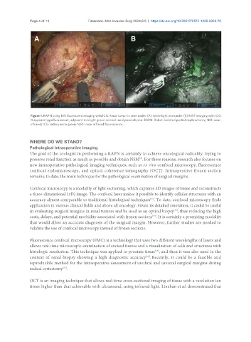
Figure 1. RAPN using NIR fluorescent imaging with ICG. Renal tumor is seen under (A) white light and under (B) NIRF imaging with ICG.
It appears hypofluorescent, adjacent to bright green normal renal parenchyma. RAPN: Robot-assisted partial nephrectomy; NIR: near-
infrared; ICG: indocyanine green; NIRF: near-infrared fluorescence.
WHERE DO WE STAND?
Pathological intraoperative imaging
The goal of the urologist in performing a RAPN is certainly to achieve oncological radicality, trying to
[3]
preserve renal function as much as possible and obtain NSM . For these reasons, research also focuses on
new intraoperative pathological imaging techniques, such as ex vivo confocal microscopy, fluorescence
confocal endomicroscopy, and optical coherence tomography (OCT). Intraoperative frozen section
remains, to date, the main technique for the pathological examination of surgical margins.
Confocal microscopy is a modality of light sectioning, which captures 2D images of tissue and reconstructs
a three-dimensional (3D) image. The confocal laser makes it possible to identify cellular structures with an
accuracy almost comparable to traditional histological techniques . To date, confocal microscopy finds
[32]
application in various clinical fields and above all oncology. Given its detailed resolution, it could be useful
in evaluating surgical margins in renal tumors and be used as an optical biopsy , thus reducing the high
[33]
costs, delays, and potential morbidity associated with frozen sections . It is certainly a promising modality
[34]
that would allow an accurate diagnosis of the surgical margin. However, further studies are needed to
validate the use of confocal microscopy instead of frozen sections.
Fluorescence confocal microscopy (FMC) is a technology that uses two different wavelengths of lasers and
allows real-time microscopic examination of excised tissues and a visualization of cells and structures with
[35]
histologic resolution. This technique was applied to prostate tissue , and then it was also used in the
context of renal biopsy showing a high diagnostic accuracy . Recently, it could be a feasible and
[36]
reproducible method for the intraoperative assessment of urethral and ureteral surgical margins during
[37]
radical cystectomy .
OCT is an imaging technique that allows real-time cross-sectional imaging of tissue with a resolution ten
times higher than that achievable with ultrasound, using infrared light. Linehan et al. demonstrated that