
Page 23 - Read Online
P. 23
Page 4 of 10 De Nunzio et al. Mini-invasive Surg 2024;8:22 https://dx.doi.org/10.20517/2574-1225.2023.138
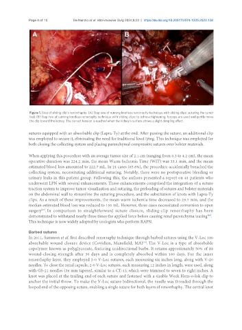
Figure 1. Step of sliding clip’s renorrhaphy. (A) Step one of running knotless renorraphy technique with sliding clips: suturing the tumor
bed; (B) Step two of running knotless renorraphy technique with sliding clips: to achieve tightening, forceps are used and gently move
the clip toward the kidney. The correct tension is reached when the kidney’s surface shows a slight dimpling effect.
sutures equipped with an absorbable clip (Lapra-Ty) at the end. After passing the suture, an additional clip
was employed to secure it, eliminating the need for traditional knot tying. This technique was employed for
both closing the collecting system and placing parenchymal compressive sutures over bolster materials.
When applying this procedure with an average tumor size of 2.1 cm (ranging from 0.3 to 4.2 cm), the mean
operative duration was 224.2 min, the mean Warm Ischemia Time (WIT) was 33.1 min, and the mean
estimated blood loss amounted to 222.7 mL. In 21 cases (65.6%), the procedure accidentally breached the
collecting system, necessitating additional suturing. Notably, there were no postoperative bleeding or
urinary leaks in this patient group. Following this, the authors presented a report on 41 patients who
underwent LPN with several enhancements. These enhancements comprised the integration of a suture
traction system to improve tumor visualization and suturing, the preloading of sutures and bolster materials
on the abdominal wall to streamline the suturing procedure, and the substitution of knots with Lapra-Ty
clips. As a result of these improvements, the mean warm ischemia time decreased to 29.7 min, and the
median estimated blood loss was reduced to 150 mL. However, three cases necessitated conversion to open
surgery . In comparison to straightforward suture closure, sliding-clip renorrhaphy has been
[27]
demonstrated to withstand nearly three times the applied force before causing renal parenchyma tearing .
[28]
This technique is now widely adopted by urologists who perform RAPN.
Barbed sutures
In 2011, Sammon et al. first described renorraphy technique through barbed sutures using the V-Loc 180
[29]
absorbable wound closure device (Covidien, Mansfield, MA) . The V-Loc is a type of absorbable
copolymer known as polyglyconate, featuring unidirectional barbs. It retains approximately 50% of its
wound-closing strength after 30 days and is completely absorbed within 180 days. For the inner
renorrhaphy layer, they employed 3-0 V-Loc sutures, each measuring six inches long, along with V-20
needles. To close the renal capsule, 2-0 V-Loc sutures, each measuring 12 inches in length, were used, along
with GS-21 needles (36 mm tapered, similar to a CT-1), which were trimmed to seven to eight inches. A
knot was placed at the trailing end of each suture and fastened with a sizable Weck Hem-o-lok clip to
anchor the initial throw. To make the V-Loc suture bidirectional, the needle was threaded through the
looped end of the opposing suture, enabling a single suture for both layers of renorrhaphy. The central knot