
Page 26 - Read Online
P. 26
De Nunzio et al. Mini-invasive Surg 2024;8:22 https://dx.doi.org/10.20517/2574-1225.2023.138 Page 7 of 10
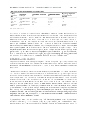
Table 1. Renal functional outcomes based on renorrhaphy technique
Authors Renorrhaphy Percentage of volume loss Percentage of GFR loss Percentage of ipsilateral
Publication technique (%) (%) split renal function
year decrease
Bahler et al. [36] Single layer vs. double 3.8 vs. 15.6 2.4 vs. 8.9 n/a
(2015) layer P < 0.001 P = 0.03
Bahler et al. [38] Single layer vs. double 9 vs. 17 4.4 vs. 8.8 n/a
(2015) layer P = 0.003 P = 0.14
Porpiglia et al. [37] Single layer vs. double n/a n/a 7.3 vs. 16.3
(2016) layer P < 0.05
GFR: Glomerular filtration rate.
documented. In most of the studies considered in this analysis, injuries to the UCS, which tend to occur
more frequently in cases involving larger and/or anatomically intricate renal masses, were managed using
different thread types. Repair methods ranged from selective interrupted sutures (including figure-of-eight
sutures) to incorporating repair within the running suture for inner-layer renorrhaphy. Desai et al.
compared UCS repair and non-repair during partial nephrectomy, and discovered that repairing UCS
injuries was linked to a significantly longer WIT . However, no discernible differences in terms of
[39]
functional outcomes or complication rates were found. Among the studies that compared running sutures
to interrupted sutures, four of them documented the use of a specialized suture for the UCS [40-43] . The
frequently utilized sutures were 2/0 or 3/0 polyglactin sutures. Williams et al. omitted the inner-layer suture
in their study . In particular, the authors performed a dedicated closure of the UCS and closed the outer
[44]
layer by using sliding-clip renorrhaphy. Such kind of suture did not increase the risk of complications and
did not reduce the renal function when compared to the double-layer renorrhaphy technique .
[21]
DISCUSSION AND CONCLUSION
Important data support the idea that preserving renal function after partial nephrectomy involves using
complex techniques that avoid indiscriminate and compressive stitching of the renal parenchyma. Several
studies have suggested some technical refinements in RPN to make intracorporeal suturing easier and
reduce WIT.
The Cleveland Clinic Group introduced an early unclamping method that led to a notable reduction in
WIT, enhanced visualization, and better management of residual bleeding during renorrhaphy. Another
noteworthy modification entailed the substitution of conventional freehand knot-tying with either a sliding-
clip technique or the use of self-retaining barbed sutures. Sliding-clip techniques were created to improve
control and tension adjustment during suturing to reduce WIT and the potential for the “cheese-cutting
effect” often associated with traditional sutures. Regardless of the specific type of suture employed, running
sutures for both the inner and outer layers of renorrhaphy have been established to minimize WIT and
simplify intracorporeal suturing during RPN. Selective suturing or a sutureless technique in RPN appears
viable and secure . Moreover, these methods represent less invasive surgical approaches. Several studies
[45]
have reported positive results regarding the accomplishment of the trifecta and functional outcomes,
confirming the safety and feasibility of this innovative and challenging technique. Additionally, when
performing renorrhaphy, the focus should be on minimizing complications related to partial nephrectomy,
such as bleeding and urinary fistulas [Table 2].
During medullary renorrhaphy, it is crucial to avoid overly deep needle passages to prevent the involvement
of the UCS and minimize the risk of vascular fistulas. In the event of a UCS injury occurring during tumor
excision, accurate superficial sutures or individual absorbable clips can be effectively employed to close the