
Page 13 - Read Online
P. 13
Page 6 of 12 Pecoraro et al. Mini-invasive Surg 2024;8:25 https://dx.doi.org/10.20517/2574-1225.2023.134
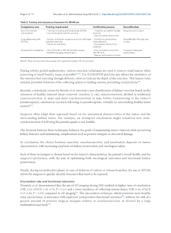
Table 3. Training and competency framework for 3DVM use
Competency area Training requirement Certification process Recertification
Basic 3D anatomy - Training on reading and interpreting 3DVMs - Complete accredited training Required every 2 years
interpretation - Use of simulation tools for practice modules
- Pass simulation-based test
Surgical planning with - Specific training for surgeons on tumor and organ - Certified by professional Recertification through case
3DVMs resection planning medical boards studies
- Hands-on workshops - Assessment during mock
surgeries
Intraoperative navigation - Use of 3DVMs in AR/VR-assisted surgery - Pass competency test with Ongoing training and
- Real-time imaging interpretation AR/VR tools assessments
- Regular performance reviews
3DVM: Three-dimensional virtual model; AR: augmented reality; VR: virtual reality.
During robotic partial nephrectomy, various resection techniques are used to remove renal tumors while
preserving as much healthy tissue as possible [26-28] . The ICON3DTM platform also allows the simulation of
the resection bed reporting through different colors to indicate the depth of the resection. This feature helps
[18]
simulate potential violations of the collecting system or feeding arteries, preventing complications .
Recently, a systematic review by Bertolo et al. reported a new classification of kidney resection based on the
extension of healthy tumoral tissue removed: resection (1 cm), enucleoresection, divided in traditional
enucleoresection (2 mm) and mini-enucleoresection (2 mm, before transitioning to the tumor’s
pseudocapsule), enucleation (excision following its pseudocapsule, virtually no surrounding healthy tissue
excised) .
[26]
Surgeons often adapt their approach based on the anatomical characteristics of the tumor and the
surrounding kidney tissue. For example, an attempted enucleation might transition into mini-
enucleoresection if following the pseudocapsule is not feasible.
The decision between these techniques balances the goals of maximizing tumor removal while preserving
kidney function and minimizing complications such as positive margins or structural damage.
In conclusion, the choice between resection, enucleoresection, and enucleation depends on tumor
characteristics, with increasing emphasis on kidney preservation and oncological safety.
Each of these techniques is chosen based on the tumor’s characteristics, the patient’s overall health, and the
surgeon’s preference, with the aim of optimizing both oncological outcomes and functional kidney
preservation.
Finally, during reconstructive phase, in case of violation of calyces or venous branches, the use of 3DVMs
allows the surgeon to quickly identify structures that need to be repaired.
Enucleation rate and functional outcomes
Piramide et al. demonstrated that the use of 3D imaging during NSS resulted in higher rates of enucleation
(OR: 2.54, 95%CI: 1.36-4.74; P < 0.01) and a lower incidence of collecting system injury (OR: 0.36, 95%CI:
[23]
0.15-0.89; P = 0.03) compared to 2D imaging . The enucleation technique, which preserves more healthy
renal parenchyma, is associated with improved postoperative functional outcomes , without the risk of a
[27]
greater amount of positive surgical margins relative to enucleoresection, as showed by a large
multinstitutional study .
[28]