
Page 10 - Read Online
P. 10
Cho et al. Mini-invasive Surg 2021;5:20 https://dx.doi.org/10.20517/2574-1225.2021.11 Page 3 of 8
Figure 1. Endoscopic radiofrequency electrode and power generator (STARmed, Koyang, Korea): 19-gauge endoscopic radiofrequency
ablation electrode (A); and a VIVA radiofrequency power generator (B).
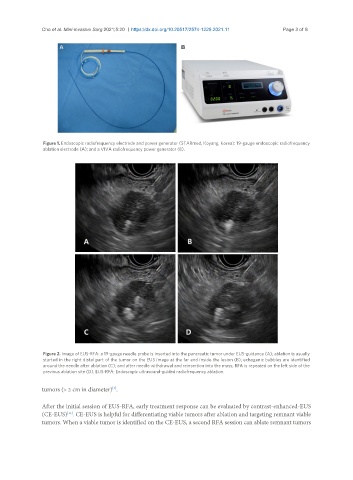
Figure 2. Image of EUS-RFA: a 19-gauge needle probe is inserted into the pancreatic tumor under EUS-guidance (A); ablation is usually
started in the right distal part of the tumor on the EUS image at the far end inside the lesion (B); echogenic bubbles are identified
around the needle after ablation (C); and after needle withdrawal and reinsertion into the mass, RFA is repeated on the left side of the
previous ablation site (D). EUS-RFA: Endoscopic ultrasound-guided radiofrequency ablation.
[5]
tumors (> 2 cm in diameter) .
After the initial session of EUS-RFA, early treatment response can be evaluated by contrast-enhanced-EUS
[11]
(CE-EUS) . CE-EUS is helpful for differentiating viable tumors after ablation and targeting remnant viable
tumors. When a viable tumor is identified on the CE-EUS, a second RFA session can ablate remnant tumors