
Page 14 - Read Online
P. 14
Page 6 of 8 Cho et al. Mini-invasive Surg 2021;5:20 https://dx.doi.org/10.20517/2574-1225.2021.11
Figure 3. Contrast-enhanced-EUS showing a nonenhanced necrotic portion (arrow) with an enhanced viable tumor. EUS: Endoscopic
ultrasound.
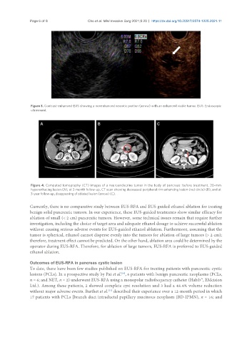
Figure 4. Computed tomography (CT) images of a neuroendocrine tumor in the body of pancreas: before treatment, 20-mm
hyperenhacing lesion (A); at 3-month follow-up, CT scan showing decreased peripheral rim enhancing lesion (red circle) (B); and at
3-year follow-up, disappearing of ablated lesion (arrow) (C).
Currently, there is no comparative study between EUS-RFA and EUS-guided ethanol ablation for treating
benign solid pancreatic tumors. In our experience, these EUS-guided treatments show similar efficacy for
ablation of small (< 2 cm) pancreatic tumors. However, some technical issues remain that require further
investigation, including the choice of target area and adequate ethanol dosage to achieve successful ablation
without causing serious adverse events for EUS-guided ethanol ablation. Furthermore, assuming that the
tumor is spherical, ethanol cannot disperse evenly into the tumors for ablation of large tumors (> 2 cm);
therefore, treatment effect cannot be predicted. On the other hand, ablation area could be determined by the
operator during EUS-RFA. Therefore, for ablation of large tumors, EUS-RFA is preferred to EUS-guided
ethanol ablation.
Outcomes of EUS-RFA in pancreas cystic lesion
To date, there have been few studies published on EUS-RFA for treating patients with pancreatic cystic
lesions (PCLs). In a prospective study by Pai et al. , 6 patients with benign pancreatic neoplasms (PCLs,
[19]
n = 6; and NET, n = 2) underwent EUS-RFA using a monopolar radiofrequency catheter (Habib™, EMcision
Ltd.). Among these patients, 2 showed complete cyst resolution and 3 had a 48.4% volume reduction
without major adverse events. Barthet et al. described their experience over a 12-month period in which
[13]
17 patients with PCLs [branch duct intraductal papillary mucinous neoplasm (BD-IPMN), n = 16; and