
Page 43 - Read Online
P. 43
Page 6 of 22 Gharagozloo et al. Mini-invasive Surg 2020;4:68 I http://dx.doi.org/10.20517/2574-1225.2020.60
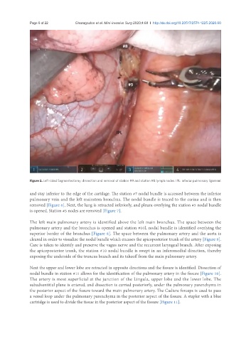
Figure 5. Left sided Segmentectomy: dissection and removal of station #9 and station #8 lymph nodes. IPL: inferior pulmonary ligament
and stay inferior to the edge of the cartilage. The station #7 nodal bundle is accessed between the inferior
pulmonary vein and the left mainstem bronchus. The nodal bundle is traced to the carina and is then
removed [Figure 6]. Next, the lung is retracted inferiorly, and pleura overlying the station #5 nodal bundle
is opened. Station #5 nodes are removed [Figure 7].
The left main pulmonary artery is identified above the left main bronchus. The space between the
pulmonary artery and the bronchus is opened and station #10L nodal bundle is identified overlying the
superior border of the bronchus [Figure 8]. The space between the pulmonary artery and the aorta is
cleared in order to visualize the nodal bundle which encases the apicoposterior trunk of the artery [Figure 9].
Care is taken to identify and preserve the vagus nerve and the recurrent laryngeal branch. After exposing
the apicoposterior trunk, the station #10 nodal bundle is swept in an inferomedial direction, thereby
exposing the underside of the truncus branch and its takeoff from the main pulmonary artery.
Next the upper and lower lobe are retracted in opposite directions and the fissure is identified. Dissection of
nodal bundle in station #11 allows for the identification of the pulmonary artery in the fissure [Figure 10].
The artery is most superficial at the junction of the Lingula, upper lobe and the lower lobe. The
subadventitial plane is entered, and dissection is carried posteriorly, under the pulmonary parenchyma in
the posterior aspect of the fissure toward the main pulmonary artery. The Cadiere forceps is used to pass
a vessel loop under the pulmonary parenchyma in the posterior aspect of the fissure. A stapler with a blue
cartridge is used to divide the tissue in the posterior aspect of the fissure [Figure 11].