
Page 40 - Read Online
P. 40
Gharagozloo et al. Mini-invasive Surg 2020;4:68 I http://dx.doi.org/10.20517/2574-1225.2020.60 Page 3 of 22
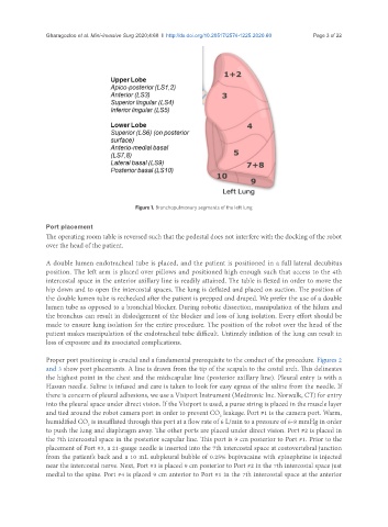
Figure 1. Bronchopulmonary segments of the left lung
Port placement
The operating room table is reversed such that the pedestal does not interfere with the docking of the robot
over the head of the patient.
A double lumen endotracheal tube is placed, and the patient is positioned in a full lateral decubitus
position. The left arm is placed over pillows and positioned high enough such that access to the 4th
intercostal space in the anterior axillary line is readily attained. The table is flexed in order to move the
hip down and to open the intercostal spaces. The lung is deflated and placed on suction. The position of
the double lumen tube is rechecked after the patient is prepped and draped. We prefer the use of a double
lumen tube as opposed to a bronchial blocker. During robotic dissection, manipulation of the hilum and
the bronchus can result in dislodgement of the blocker and loss of lung isolation. Every effort should be
made to ensure lung isolation for the entire procedure. The position of the robot over the head of the
patient makes manipulation of the endotracheal tube difficult. Untimely inflation of the lung can result in
loss of exposure and its associated complications.
Proper port positioning is crucial and a fundamental prerequisite to the conduct of the procedure. Figures 2
and 3 show port placements. A line is drawn from the tip of the scapula to the costal arch. This delineates
the highest point in the chest and the midscapular line (posterior axillary line). Pleural entry is with a
Hassan needle. Saline is infused and care is taken to look for easy egress of the saline from the needle. If
there is concern of pleural adhesions, we use a Visiport Instrument (Medtronic Inc. Norwalk, CT) for entry
into the pleural space under direct vision. If the Visiport is used, a purse string is placed in the muscle layer
and tied around the robot camera port in order to prevent CO leakage. Port #1 is the camera port. Warm,
2
humidified CO is insufflated through this port at a flow rate of 6 L/min to a pressure of 6-8 mmHg in order
2
to push the lung and diaphragm away. The other ports are placed under direct vision. Port #2 is placed in
the 7th intercostal space in the posterior scapular line. This port is 9 cm posterior to Port #1. Prior to the
placement of Port #3, a 21-gauge needle is inserted into the 7th intercostal space at costovertebral junction
from the patient’s back and a 10 mL subpleural bubble of 0.25% bupivacaine with epinephrine is injected
near the intercostal nerve. Next, Port #3 is placed 9 cm posterior to Port #2 in the 7th intercostal space just
medial to the spine. Port #4 is placed 9 cm anterior to Port #1 in the 7th intercostal space at the anterior