
Page 47 - Read Online
P. 47
Page 10 of 22 Gharagozloo et al. Mini-invasive Surg 2020;4:68 I http://dx.doi.org/10.20517/2574-1225.2020.60
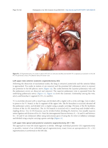
Figure 12. LS3 Segmentectomy: the nodes in station #5 (LN) are removed and the proximal left PA is exposed just posterior to the left
PN. PA: pulmonary artery; PN: phrenic nerve; LN: lymph node
Left upper lobe anterior anatomic segmentectomy (S3)
Following the dissection of mediastinal nodes, the lung is retracted posteriorly and the anterior hilum
is approached. The nodes in station #5 are removed and the proximal left pulmonary artery is exposed
just posterior to the left phrenic nerve [Figure 12]. The nodes between the superior pulmonary vein and
the pulmonary artery are dissected and removed. The superior pulmonary vein is separated from the
underlying pulmonary artery [Figure 13]. Figure 14 shows the anatomic relationship among the vein,
artery, and bronchus in segment S3 (V3, A3 and B3).
V3 is encircled, elevated with a vessel loop, and divided with a stapler with a white cartridge. Care is taken
to preserve the V1 branch to the S1 segment of the upper lobe. The B3 bronchus is encircled, elevated off
the pulmonary artery, and divided with a stapler using a purple cartridge. Division of the B3 facilitates
division of the A3 PA branch(es). The A3 PA branch is encircled with a vessel loop and divided with a
stapling device. The A3 PA branches can be divided before dividing B3; however, this usually requires
suture ligation and division of the A3. Next the intersegmental fissures between S1 + S2 and S3 and between
S4 + S5 and S3 are delineated either using indocyanine green if using the Xi robot or inflation technique
and divided using a stapler carrying a green cartridge [Figure 15].
Left upper lobe apical and posterior anatomic segmentectomy (S1 + S2)
The approach to these left sided segments is similar. Although individual posterior (S2) segmentectomy
is possible, instead of an individual apical segmentectomy, many times an apicoposterior (S1 + S2)
segmentectomy is performed on the left side.