
Page 49 - Read Online
P. 49
Page 12 of 22 Gharagozloo et al. Mini-invasive Surg 2020;4:68 I http://dx.doi.org/10.20517/2574-1225.2020.60
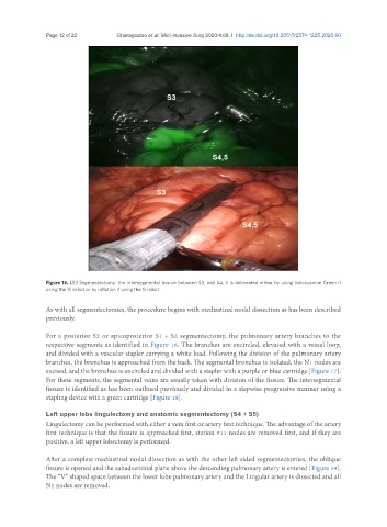
Figure 15. LS3 Segmentectomy: the intersegmental fissure between S3, and S4, 5 is delineated either by using Indocyanine Green if
using the Xi robot or by inflation if using the Si robot
As with all segmentectomies, the procedure begins with mediastinal nodal dissection as has been described
previously.
For a posterior S2 or apicoposterior S1 + S2 segmentectomy, the pulmonary artery branches to the
respective segments as identified in Figure 16. The branches are encircled, elevated with a vessel loop,
and divided with a vascular stapler carrying a white load. Following the division of the pulmonary artery
branches, the bronchus is approached from the back. The segmental bronchus is isolated, the N1 nodes are
excised, and the bronchus is encircled and divided with a stapler with a purple or blue cartridge [Figure 17].
For these segments, the segmental veins are usually taken with division of the fissure. The intersegmental
fissure is identified as has been outlined previously and divided in a stepwise progressive manner using a
stapling device with a green cartridge [Figure 18].
Left upper lobe lingulectomy and anatomic segmentectomy (S4 + S5)
Lingulectomy can be performed with either a vein first or artery first technique. The advantage of the artery
first technique is that the fissure is approached first, station #11 nodes are removed first, and if they are
positive, a left upper lobectomy is performed.
After a complete mediastinal nodal dissection as with the other left sided segmentectomies, the oblique
fissure is opened and the subadventitial plane above the descending pulmonary artery is entered [Figure 19].
The “V” shaped space between the lower lobe pulmonary artery and the Lingular artery is dissected and all
N1 nodes are removed.