
Page 54 - Read Online
P. 54
Gharagozloo et al. Mini-invasive Surg 2020;4:68 I http://dx.doi.org/10.20517/2574-1225.2020.60 Page 17 of 22
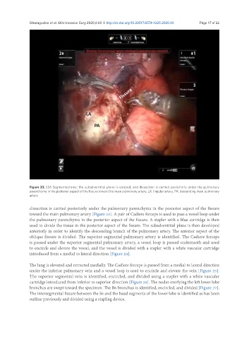
Figure 23. LS6 Segmentectomy: the subadventitial plane is entered, and dissection is carried posteriorly under the pulmonary
parenchyma in the posterior aspect of the fissure toward the main pulmonary artery. LA: lingular artery; PA: descending main pulmonary
artery
dissection is carried posteriorly under the pulmonary parenchyma in the posterior aspect of the fissure
toward the main pulmonary artery [Figure 23]. A pair of Cadiere forceps is used to pass a vessel loop under
the pulmonary parenchyma in the posterior aspect of the fissure. A stapler with a blue cartridge is then
used to divide the tissue in the posterior aspect of the fissure. The subadventitial plane is then developed
anteriorly in order to identify the descending branch of the pulmonary artery. The anterior aspect of the
oblique fissure is divided. The superior segmental pulmonary artery is identified. The Cadiere forceps
is passed under the superior segmental pulmonary artery, a vessel loop is passed underneath and used
to encircle and elevate the vessel, and the vessel is divided with a stapler with a white vascular cartridge
introduced from a medial to lateral direction [Figure 24].
The lung is elevated and retracted medially. The Cadiere forceps is passed from a medial to lateral direction
under the inferior pulmonary vein and a vessel loop is used to encircle and elevate the vein [Figure 25].
The superior segmental vein is identified, encircled, and divided using a stapler with a white vascular
cartridge introduced from inferior to superior direction [Figure 26]. The nodes overlying the left lower lobe
bronchus are swept toward the specimen. The B6 bronchus is identified, encircled, and divided [Figure 27].
The intersegmental fissure between the S6 and the basal segments of the lower lobe is identified as has been
outline previously and divided using a stapling device.