
Page 58 - Read Online
P. 58
Shiozaki et al. Mini-invasive Surg 2020;4:50 I http://dx.doi.org/10.20517/2574-1225.2020.31 Page 5 of 9
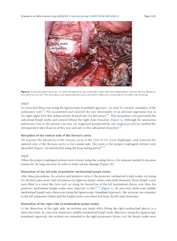
Figure 4. Anomalous pulmonary vein. An aberrant segmental vein in the right upper lobe that independently drained into the left atrium
was identified (arrow). This anomalous vein penetrated the subcarinal lymph nodes and crossed behind the right main bronchus
Pitfall
In subcarinal dissection using the laparoscopic transhiatal approach, we need to consider anomalies of the
[18]
pulmonary vein . We encountered and reported the rare abnormality of an aberrant segmental vein in
[18]
the right upper lobe that independently drained into the left atrium . This anomalous vein penetrated the
subcarinal lymph nodes and crossed behind the right main bronchus [Figure 4]. Although the anomalous
pulmonary vein in the present case was not diagnosed preoperatively, our surgical procedure enabled the
[18]
intraoperative identification of this vein and safe en bloc subcarinal dissection .
Abruption of the ventral side of the thoracic aorta
We exposed the adventitia of the thoracic aorta at the level of the crural diaphragm, and dissected the
anterior side of the thoracic aorta to the cranial side. The roots of the proper esophageal arteries were
identified [Figure 5A] and divided using the long sealing device [8-12] .
Pitfall
When the proper esophageal arteries were divided using the sealing device, the assistant needed to decrease
tension by the long retractor in order to avoid arterial damage [Figure 5B].
Dissection of the left side of posterior mediastinal lymph nodes
After these procedures, the anterior and posterior sides of the posterior mediastinal lymph nodes, including
the thoracic para-aortic and left pulmonary ligament lymph nodes, were both dissected. These lymph nodes
were lifted in a sheet-like form and cut along the borderline of the left mediastinal pleura, and, thus, the
posterior mediastinal lymph nodes were dissected en bloc [8-12] [Figure 6]. In cases that underwent middle
mediastinal lymph node dissection using the laparoscopic transhiatal approach, this incision was extended
to the left pulmonary hilum and the lymph nodes were dissected from the left main bronchus.
Dissection of the right side of mediastinal lymph nodes
In the dissection of the right side, an incision was made while lifting the right mediastinal pleura in a
sheet-like form. In cases that underwent middle mediastinal lymph node dissection using the laparoscopic
transhiatal approach, the incision was extended to the right pulmonary hilum, and the lymph nodes were