
Page 56 - Read Online
P. 56
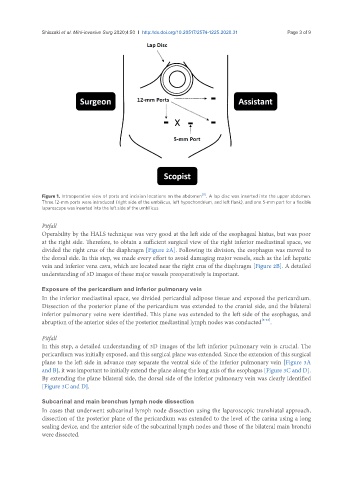
Shiozaki et al. Mini-invasive Surg 2020;4:50  I  http://dx.doi.org/10.20517/2574-1225.2020.31                                  Page 3 of 9


                                                                     [11]
               Figure 1. Intraoperative view of ports and incision locations on the abdomen . A lap disc was inserted into the upper abdomen.
               Three 12-mm ports were introduced (right side of the umbilicus, left hypochondrium, and left flank), and one 5-mm port for a flexible
               laparoscope was inserted into the left side of the umbilicus
               Pitfall
               Operability by the HALS technique was very good at the left side of the esophageal hiatus, but was poor
               at the right side. Therefore, to obtain a sufficient surgical view of the right inferior mediastinal space, we
               divided the right crus of the diaphragm [Figure 2A]. Following its division, the esophagus was moved to
               the dorsal side. In this step, we made every effort to avoid damaging major vessels, such as the left hepatic
               vein and inferior vena cava, which are located near the right crus of the diaphragm [Figure 2B]. A detailed
               understanding of 3D images of these major vessels preoperatively is important.

               Exposure of the pericardium and inferior pulmonary vein
               In the inferior mediastinal space, we divided pericardial adipose tissue and exposed the pericardium.
               Dissection of the posterior plane of the pericardium was extended to the cranial side, and the bilateral
               inferior pulmonary veins were identified. This plane was extended to the left side of the esophagus, and
               abruption of the anterior sides of the posterior mediastinal lymph nodes was conducted [8-12] .

               Pitfall
               In this step, a detailed understanding of 3D images of the left inferior pulmonary vein is crucial. The
               pericardium was initially exposed, and this surgical plane was extended. Since the extension of this surgical
               plane to the left side in advance may separate the ventral side of the inferior pulmonary vein [Figure 3A
               and B], it was important to initially extend the plane along the long axis of the esophagus [Figure 3C and D].
               By extending the plane bilateral side, the dorsal side of the inferior pulmonary vein was clearly identified
               [Figure 3C and D].


               Subcarinal and main bronchus lymph node dissection
               In cases that underwent subcarinal lymph node dissection using the laparoscopic transhiatal approach,
               dissection of the posterior plane of the pericardium was extended to the level of the carina using a long
               sealing device, and the anterior side of the subcarinal lymph nodes and those of the bilateral main bronchi
               were dissected.