
Page 57 - Read Online
P. 57
Page 4 of 9 Shiozaki et al. Mini-invasive Surg 2020;4:50 I http://dx.doi.org/10.20517/2574-1225.2020.31
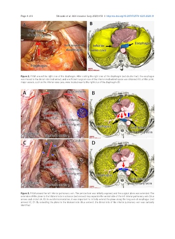
Figure 2. Pitfall around the right crus of the diaphragm. After cutting the right crus of the diaphragm (red double line), the esophagus
was moved to the dorsal side (red arrow), and a sufficient surgical view of the inferior mediastinal space was obtained (A); at this point,
major vessels, such as the inferior vena cava, were located near to the right crus of the diaphragm (B)
Figure 3. Pitfall around the left inferior pulmonary vein. The pericardium was initially exposed, and the surgical plane was extended. The
extension of this plane to the bilateral side in advance (red arrows) may separate the ventral side of the left inferior pulmonary vein (blue
arrows and circle) (A, B); to avoid misorientation, it was important to initially extend the plane along the long axis of esophagus (red
arrows) (C, D). By extending the plane to the bilateral side (blue arrows), the dorsal side of the inferior pulmonary vein was certainly
identified