
Page 27 - Read Online
P. 27
Page 4 of 9 Shirakawa et al. Mini-invasive Surg 2020;4:33 I http://dx.doi.org/10.20517/2574-1225.2020.30
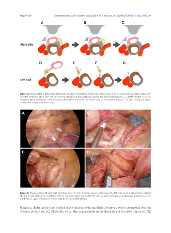
Figure 2. Microanatomy-based standardization of upper mediastinal lymph node dissection. A, D: detaching the esophagus together
with the lymphatic chain from the trachea and aggregating the lymphatic chain to the esophageal side; B, E, F: identifying the recurrent
laryngeal nerve and lymph node dissection along the nerve from the central part to the peripheral part; C, G: final findings of upper
mediastinal lymph node dissection
A B
C D
Figure 3. Thoracoscopic 4K ultra-high-definition view. A: detaching the meso-esophagus on the left side while preserving the visceral
sheath; B: aggregating the lymphatic chain to the esophageal side on the left side; C: upper mediastinal lymph node dissection on the
right side; D: upper mediastinal lymph node dissection on the left side
lymphatic chain on the inner surface of the visceral sheath and slide the nerve down to the natural position
[Figures 2B, E, F and 3C, D]. Finally, we cut the visceral sheath on the dorsal side of the nerve [Figure 2C, G].