
Page 26 - Read Online
P. 26
Shirakawa et al. Mini-invasive Surg 2020;4:33 I http://dx.doi.org/10.20517/2574-1225.2020.30 Page 3 of 9
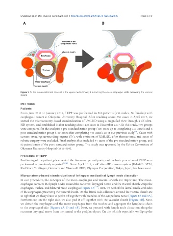
Figure 1. A: the microanatomical concept in the upper mediastinum; B: detaching the meso-esophagus while preserving the visceral
sheath
METHODS
Patients
From June 2011 to January 2019, TEPP was performed in 500 patients (430 males, 70 females) with
esophageal cancer at Okayama University Hospital. After reaching about 350 cases in April 2017, we
started the microanatomy-based standardization of UMLND using a magnified view through a 4K ultra-
HD system, and established it after reaching about 400 cases in November 2017. In this study, two groups
were compared for the analysis: a pre-standardization group (100 cases up to completing 350 cases) and a
[19]
post-standardization group (100 cases after completing 400 cases), as in our previous study . Cases with
tumors invading surrounding organs (T4), with omission of UMLND, after thoracotomy, and cases of
robotic surgery were excluded. Final analysis thus included 91 cases of the pre-standardization group, and
83 paired cases of the post-standardization group. This study was approved by the Ethics Committee of
Okayama University Hospital (1811-009).
Procedure of TEPP
Positioning of the patient, placement of the thoracoscope and ports, and the basic procedure of TEPP were
performed as previously reported [19,20] . Since April 2017, a 4K ultra-HD camera system (IMAGE1 STM,
Karl Storz, Tuttlingen, Germany and Visera 4K UHD, Olympus Corporation, Tokyo, Japan) has been used.
Microanatomy-based standardization of left upper mediastinal lymph node dissection
In our procedure, the concepts of the meso-esophagus and visceral sheath are important. The meso-
esophagus contains the lymph nodes around the recurrent laryngeal nerve, and the visceral sheath wraps the
[19]
esophagus, trachea, and bilateral meso-esophagus [Figure 1A] . First, we peel off the dorsal and lateral sides
of the esophagus, preserving the visceral sheath. On the lateral side, adhesions around the visceral sheath are
so tight that we always have to peel it off together with branches of the sympathetic nerve [Figure 1B and 3A].
Furthermore, on the right side, we also peel it off together with the vascular sheath [Figure 1B]. Next,
we detach the esophagus and the meso-esophagus from the trachea and aggregate the lymphatic chain
to the esophageal side [Figures 2A, D and 3B]. Next, we proceed with lymph node dissection along the
recurrent laryngeal nerve from the central to the peripheral part. On the left side especially, we flip up the