
Page 78 - Read Online
P. 78
Page 4 of 6 Abu Akar et al. Mini-invasive Surg 2020;4:25 I http://dx.doi.org/10.20517/2574-1225.2019.66
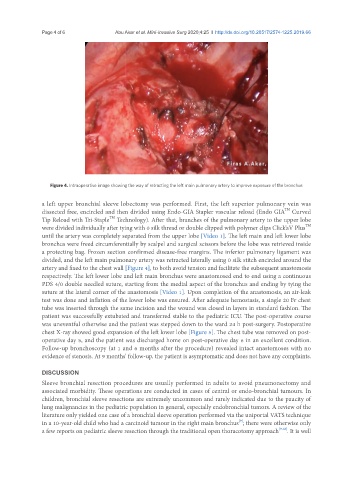
Figure 4. Intraoperative image showing the way of retracting the left main pulmonary artery to improve exposure of the bronchus
a left upper bronchial sleeve lobectomy was performed. First, the left superior pulmonary vein was
TM
dissected free, encircled and then divided using Endo-GIA Stapler vascular reload (Endo GIA Curved
TM
Tip Reload with Tri-Staple Technology). After that, branches of the pulmonary artery to the upper lobe
TM
were divided individually after tying with 0 silk thread or double clipped with polymer clips Click’aV Plus
until the artery was completely separated from the upper lobe [Video 1]. The left main and left lower lobe
bronchus were freed circumferentially by scalpel and surgical scissors before the lobe was retrieved inside
a protecting bag. Frozen section confirmed disease-free margins. The inferior pulmonary ligament was
divided, and the left main pulmonary artery was retracted laterally using 0 silk stitch encircled around the
artery and fixed to the chest wall [Figure 4], to both avoid tension and facilitate the subsequent anastomosis
respectively. The left lower lobe and left main bronchus were anastomosed end to end using a continuous
PDS 4/0 double needled suture, starting from the medial aspect of the bronchus and ending by tying the
suture at the lateral corner of the anastomosis [Video 1]. Upon completion of the anastomosis, an air-leak
test was done and inflation of the lower lobe was ensured. After adequate hemostasis, a single 20 Fr chest
tube was inserted through the same incision and the wound was closed in layers in standard fashion. The
patient was successfully extubated and transferred stable to the pediatric ICU. The post-operative course
was uneventful otherwise and the patient was stepped down to the ward 24 h post-surgery. Postoperative
chest X-ray showed good expansion of the left lower lobe [Figure 5]. The chest tube was removed on post-
operative day 5, and the patient was discharged home on post-operative day 6 in an excellent condition.
Follow-up bronchoscopy (at 1 and 6 months after the procedure) revealed intact anastomoses with no
evidence of stenosis. At 9 months’ follow-up, the patient is asymptomatic and does not have any complaints.
DISCUSSION
Sleeve bronchial resection procedures are usually performed in adults to avoid pneumonectomy and
associated morbidity. These operations are conducted in cases of central or endo-bronchial tumours. In
children, bronchial sleeve resections are extremely uncommon and rarely indicated due to the paucity of
lung malignancies in the pediatric population in general, especially endobronchial tumors. A review of the
literature only yielded one case of a bronchial sleeve operation performed via the uniportal VATS technique
in a 10-year-old child who had a carcinoid tumour in the right main bronchus ; there were otherwise only
[9]
a few reports on pediatric sleeve resection through the traditional open thoracotomy approach [9,12] . It is well