
Page 77 - Read Online
P. 77
Abu Akar et al. Mini-invasive Surg 2020;4:25 I http://dx.doi.org/10.20517/2574-1225.2019.66 Page 3 of 6
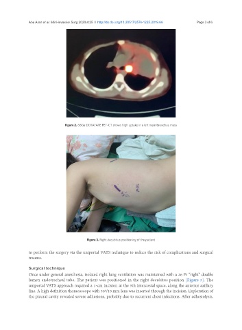
Figure 2. 68Ga DOTATATE PET-CT shows high uptake in a left main bronchus mass
Figure 3. Right decubitus positioning of the patient
to perform the surgery via the uniportal VATS technique to reduce the risk of complications and surgical
trauma.
Surgical technique
Once under general anesthesia, isolated right lung ventilation was maintained with a 26 Fr “right” double
lumen endotracheal tube. The patient was positioned in the right decubitus position [Figure 3]. The
uniportal VATS approach required a 3-cm incision at the 5th intercostal space, along the anterior axillary
line. A high definition thoracoscope with 30º/10 mm lens was inserted through the incision. Exploration of
the pleural cavity revealed severe adhesions, probably due to recurrent chest infections. After adhesiolysis,