
Page 71 - Read Online
P. 71
Page 6 of 9 Gharagozloo et al. Mini-invasive Surg 2020;4:22 I http://dx.doi.org/10.20517/2574-1225.2019.61
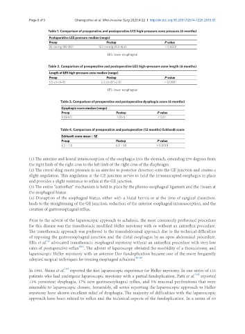
Table 1. Comparison of preoperative and postoperative LES high-pressure zone pressure (6 months)
Postoperative LES pressure median (range)
Preop Postop P value
35 mmHg (18-120) 13.2 mmHg (9.8-16.6) < 0.0001
LES: lower esophageal
Table 2. Comparison of preoperative and postoperative LES high-pressure zone length (6 months)
Length of LES high-pressure zone median (range)
Preop Postop P value
5.5 cm (4-9) 2.2 cm (1.5-2.8) < 0.0001
LES: lower esophageal
Table 3. Comparison of preoperative and postoperative dysphagia score (6 months)
Dysphagia score median (range)
Preop Postop P value
9 (8-10) 1 (0-1) < 0.01
Table 4. Comparison of preoperative and postoperative (12 months) Eckhardt score
Eckhardt score mean ± SE
Preop Postop P value
6.3 ± 1.8 0.8 ± 1.8 < 0.0001
(1) The anterior and lateral intussusception of the esophagus into the stomach, extending 270 degrees from
the right limb of the right crus to the left limb of the right crus of the diaphragm.
(2) The crural sling exerts pressure in an anterior to posterior direction onto the GE junction and creates a
slight angulation. This angulation at the GE junction serves to hold the intussuscepted esophagus in place
and provides a slight resistance to reflux at the GE junction.
(3) The entire “antireflux” mechanism is held in place by the phreno-esophageal ligament and the tissues at
the esophageal hiatus.
(4) Disruption of the esophageal hiatus, either with a hiatal hernia or at the time of surgical dissection,
leads to the straightening of the GE junction, reduction of the anterior esophageal intussusception, and the
creation of gastroesophageal reflux.
Prior to the advent of the laparoscopic approach to achalasia, the most commonly performed procedure
for this disease was the transthoracic modified Heller myotomy with or without an antireflux procedure.
The transthoracic approach was preferred to the transabdominal approach due to the technical difficulties
of exposing the gastroesophageal junction and the distal esophagus by an open abdominal procedure.
[4]
Ellis et al. advocated transthoracic esophageal myotomy without an antireflux procedure with very low
[8,9]
rates of postoperative reflux . The advent of laparoscopy obviated the morbidity of a thoracotomy, and
laparoscopic Heller myotomy with an anterior Dor fundoplication became one of the more frequently
adopted surgical techniques for treating esophageal achalasia [10-18] .
[19]
In 1991, Shimi et al. reported the first laparoscopic experience for Heller myotomy. In one series of 133
[20]
patients who had undergone laparoscopic myotomy with a partial fundoplication, Patti et al. reported
11% persistent dysphagia, 17% new gastroesophageal reflux, and 5% mucosal perforations that were
amenable to laparoscopic closure. Invariably, all series reporting the laparoscopic approach to Heller
myotomy have shown excellent relief of dysphagia. The majority of difficulties with the laparoscopic
approach have been related to reflux and the technical aspects of the fundoplication. In a series of 69