
Page 68 - Read Online
P. 68
Gharagozloo et al. Mini-invasive Surg 2020;4:22 I http://dx.doi.org/10.20517/2574-1225.2019.61 Page 3 of 9
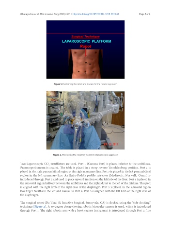
Figure 1. Positioning the robot and trocars for the robotic approach
Figure 2. Positioning the robot for the robotic laparoscopic approach
Two Laparoscopic CO insufflators are used. Port 1 (Camera Port) is placed inferior to the umbilicus.
2
Pneumoperitoneum is created. The table is placed in a steep reverse Trendelenberg position. Port 2 is
placed in the right paraumbilical region at the right mammary line. Port 3 is placed in the left paraumbilical
region in the left mammary line. An Endo-Paddle paddle retractor (Medtronic, Norwalk, Conn.) is
introduced through Port 2 and used to place upward traction on the left lobe of the liver. Port 4 is placed in
the subcostal region halfway between the umbilicus and the xiphoid just to the left of the midline. This port
is aligned with the right limb of the right crus of the diaphragm. Port 5 is placed in the subcostal region
two finger-breaths to the left and caudad to Port 4. Port 5 is aligned with the left limb of the right crus of
the diaphragm.
The surgical robot (Da Vinci Si, Intuitive Surgical, Sunnyvale, CA) is docked using the “side docking”
technique [Figure 2]. A 30-degree down-viewing robotic binocular camera is used, which is introduced
through Port 1. The right robotic arm with a hook cautery instrument is introduced through Port 3. The