
Page 69 - Read Online
P. 69
Page 4 of 9 Gharagozloo et al. Mini-invasive Surg 2020;4:22 I http://dx.doi.org/10.20517/2574-1225.2019.61
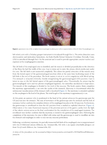
Figure 3. Laparoscopic view of the completed lateral esophageal myotomy prior to the re-approximation of the left limb of the esophageal crus
left robotic arm with a Debakey grasper instrument is introduced through Port 2. The entire dissection uses
electrocautery and meticulous hemostasis. An Endo-Paddle Retract Retractor (Covidian, Norwalk, Conn,
USA) is introduced through Port 5 by the assistant and is used to provide appropriate counter traction and
exposure at the esophagogastric junction.
The left limb of the esophageal crus is identified, and the muscle is divided perpendicular to the direction
of the fibers for half the width of the crus. Care is taken not to enter the pleura, which resides just under
the crus. The left limb is not transected completely. This allows for partial retraction of the muscle away
from the lateral aspect of the gastroesophageal junction while at the same time facilitating repair of the
left limb at the end of the procedure. The hook cautery is set at 30 cut/30 coagulation with blend setting.
The stomach is retracted inferiorly, thereby straightening the gastroesophageal (GE) junction. Care is
taken to stay on the left lateral aspect of the gastroesophageal valve. Theoretically, by preserving the
gastroesophageal valve and the phreno-esophageal ligament, the antireflux mechanism is kept intact.
The muscle of the esophagus is divided to the level of the mucosa. The hook cautery them completes
the myotomy approximately 2 cm onto the cardia of the stomach. Myotomy is discontinued when the
submucosal vascular plexus of the stomach wall is visualized [Figure 3]. The myotomy is extended cephalad
on the esophagus to the level of the pleura. The total length of the myotomy is approximately 6 cm.
At this point, an assistant who is positioned at the head of the patient advances the gastroscope past the
GE junction into the stomach. The ease of movement of the gastroscope into the stomach and the lack of
resistance further confirms the complete division of the esophageal muscles at the GE junction. Furthermore,
the gastroscope is retroflexed to view the GE junction from a caudad to cephalad direction [Figure 4].
Observation of the trans-illuminated mucosa of the proximal portion of the gastric cardia from the light
of the robotic camera serves as the final confirmation for the completion of the esophageal myotomy. The
retroflexed view further confirms that the myotomy is lateral to the gastroesophageal valve. Following the
completion of the myotomy, the area is filled with saline and the gastroscope is used to insufflate air into
the stomach and esophagus in order to rule out any mucosal perforation.
Following a satisfactory myotomy, the partially transected left limb of the esophageal crus is reapproximated
with two O-Ethibond sutures (Ethicon, Inc. Sommerville, NJ) with 2-cm square absorbable pledgets cut
from Vicryl mesh (Ethicon, Inc. Sommerville, NJ).
A video of the procedure can be accessed at https://www.youtube.com/watch?v=WUEuHSioodY&feature=
youtu.be.