
Page 61 - Read Online
P. 61
Page 6 of 10 Gharagozloo. Mini-invasive Surg 2020;4:14 I http://dx.doi.org/10.20517/2574-1225.2019.55
Sympathetic Chain
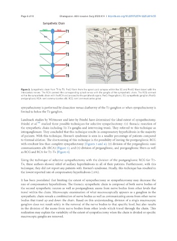
Figure 3. Sympathetic chain from T1 to T5. PreG fibers from the spinal cord synapse within the SG and PostG fibers travel with the
intercostals nerves. The RCA connect the corresponding spinal nerves with the ganglia of the sympathetic chain. The RCG connect
within the sympathetic chain with the RCA and proceed to the peripheral organs. PreG: Preganglionic; SG: sympathetic ganglion; PostG:
postganglionic; RCA: rami communicantes albi; RCG: rami communicantes grisei
sympathectomy is performed by dissection versus diathermy of the T2 ganglion or when sympathectomy is
limited to below the T2 ganglion.
Landmark studies by Wittmoser and later by Friedel have determined the ideal extent of sympathectomy.
[23]
Friedel et al. studied three possible techniques for selective sympathectomy: (1) thoracic resection of
the sympathetic chain including T2-T4 ganglia and intervening trunk. They referred to this technique as
interganglionare. They concluded that this technique results in compensatory hyperhidrosis in the majority
of patients. With this technique, Horner’s syndrome is seen in a smaller percentage of patients compared
to thermal ablation. The shortcoming of this technique is the possibility of leaving the postganglionic RCG
with resultant less than complete sympathectomy [Figures 3 and 4]; (2) division of the preganglionic rami
communicantes albi (RCA) [Figure 5]; and (3) division of preganglionic, and postganglionic fibers as well
as RCG and RCA for T2-T4 [Figure 6].
Using the technique of selective sympathectomy with the division of the postganglionic RCG for T2-
T4, these authors showed relief of axillary hyperhidrosis in all of their patients. Furthermore, with this
technique, they did not report any patients with Horner’s syndrome. Finally, this technique has resulted in
the lowest reported rate of compensatory hyperhidrosis (16%).
It has been postulated that limiting the extent of sympathectomy or sympathicotomy may decrease the
rate of compensatory hyperhidrosis. The thoracic sympathetic chain is composed of both nerve bodies of
the second sympathetic neuron as well as postganglionic axons from nerve bodies from other levels that
travel within the chain. Microscopic examination of what macroscopically appears as a ganglion in the
sympathetic chain reveals a combination of nerve bodies as well as communicating axons from other nerve
bodies that travel up and down the chain. Based on this understanding, division of a single macroscopic
ganglion does not result solely in the removal of the nerve bodies to that specific level, but also results
in the division of the axons from nerve bodies from other levels which travel through the chain. This
realization may explain the variability of the extent of sympathectomy when the chain is divided or specific
macroscopic ganglia are removed.