
Page 49 - Read Online
P. 49
Phillips et al. Mini-invasive Surg 2020;4:12 I http://dx.doi.org/10.20517/2574-1225.2020.02 Page 3 of 9
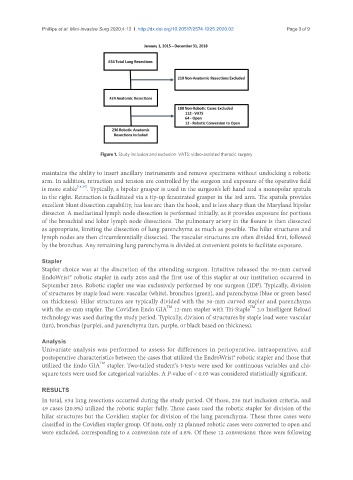
Figure 1. Study inclusion and exclusion. VATS: video-assisted thoracic surgery
maintains the ability to insert ancillary instruments and remove specimens without undocking a robotic
arm. In addition, retraction and tension are controlled by the surgeon and exposure of the operative field
is more stable [14,15] . Typically, a bipolar grasper is used in the surgeon’s left hand and a monopolar spatula
in the right. Retraction is facilitated via a tip-up fenestrated grasper in the 3rd arm. The spatula provides
excellent blunt dissection capability, has less arc than the hook, and is less sharp than the Maryland bipolar
dissector. A mediastinal lymph node dissection is performed initially, as it provides exposure for portions
of the bronchial and lobar lymph node dissections. The pulmonary artery in the fissure is then dissected
as appropriate, limiting the dissection of lung parenchyma as much as possible. The hilar structures and
lymph nodes are then circumferentially dissected. The vascular structures are often divided first, followed
by the bronchus. Any remaining lung parenchyma is divided at convenient points to facilitate exposure.
Stapler
Stapler choice was at the discretion of the attending surgeon. Intuitive released the 30-mm curved
EndoWrist® robotic stapler in early 2016 and the first use of this stapler at our institution occurred in
September 2016. Robotic stapler use was exclusively performed by one surgeon (JDP). Typically, division
of structures by staple load were: vascular (white), bronchus (green), and parenchyma (blue or green based
on thickness). Hilar structures are typically divided with the 30-mm curved stapler and parenchyma
TM
TM
with the 45-mm stapler. The Covidien Endo GIA 12-mm stapler with Tri-Staple 2.0 Intelligent Reload
technology was used during the study period. Typically, division of structures by staple load were: vascular
(tan), bronchus (purple), and parenchyma (tan, purple, or black based on thickness).
Analysis
Univariate analysis was performed to assess for differences in perioperative, intraoperative, and
postoperative characteristics between the cases that utilized the EndroWrist® robotic stapler and those that
utilized the Endo GIA stapler. Two-tailed student’s t-tests were used for continuous variables and chi-
TM
square tests were used for categorical variables. A P-value of < 0.05 was considered statistically significant.
RESULTS
In total, 634 lung resections occurred during the study period. Of those, 236 met inclusion criteria, and
49 cases (20.8%) utilized the robotic stapler fully. Three cases used the robotic stapler for division of the
hilar structures but the Covidien stapler for division of the lung parenchyma. These three cases were
classified in the Covidien stapler group. Of note, only 12 planned robotic cases were converted to open and
were excluded, corresponding to a conversion rate of 4.8%. Of these 12 conversions: three were following