
Page 44 - Read Online
P. 44
Abu Akar et al. Mini-invasive Surg 2020;4:10 I http://dx.doi.org/10.20517/2574-1225.2019.65 Page 3 of 5
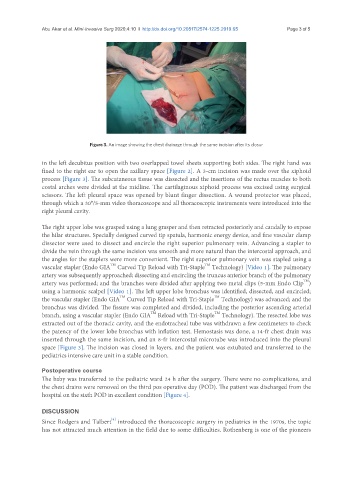
Figure 3. An image showing the chest drainage through the same incision after its closur
in the left decubitus position with two overlapped towel sheets supporting both sides. The right hand was
fixed to the right ear to open the axillary space [Figure 2]. A 3-cm incision was made over the xiphoid
process [Figure 3]. The subcutaneous tissue was dissected and the insertions of the rectus muscles to both
costal arches were divided at the midline. The cartilaginous xiphoid process was excised using surgical
scissors. The left pleural space was opened by blunt finger dissection. A wound protector was placed,
through which a 30°/5-mm video thoracoscope and all thoracoscopic instruments were introduced into the
right pleural cavity.
The right upper lobe was grasped using a lung grasper and then retracted posteriorly and caudally to expose
the hilar structures. Specially designed curved tip spatula, harmonic energy device, and fine vascular clamp
dissector were used to dissect and encircle the right superior pulmonary vein. Advancing a stapler to
divide the vein through the same incision was smooth and more natural than the intercostal approach, and
the angles for the staplers were more convenient. The right superior pulmonary vein was stapled using a
TM
TM
vascular stapler (Endo GIA Curved Tip Reload with Tri-Staple Technology) [Video 1]. The pulmonary
artery was subsequently approached; dissecting and encircling the truncus anterior branch of the pulmonary
TM
artery was performed; and the branches were divided after applying two metal clips (5-mm Endo Clip )
using a harmonic scalpel [Video 1]. The left upper lobe bronchus was identified, dissected, and encircled;
TM
TM
the vascular stapler (Endo GIA Curved Tip Reload with Tri-Staple Technology) was advanced; and the
bronchus was divided. The fissure was completed and divided, including the posterior ascending arterial
TM
TM
branch, using a vascular stapler (Endo GIA Reload with Tri-Staple Technology). The resected lobe was
extracted out of the thoracic cavity, and the endotracheal tube was withdrawn a few centimeters to check
the patency of the lower lobe bronchus with inflation test. Hemostasis was done, a 14-fr chest drain was
inserted through the same incision, and an 8-fr intercostal microtube was introduced into the pleural
space [Figure 3]. The incision was closed in layers, and the patient was extubated and transferred to the
pediatrics intensive care unit in a stable condition.
Postoperative course
The baby was transferred to the pediatric ward 24 h after the surgery. There were no complications, and
the chest drains were removed on the third pos operative day (POD). The patient was discharged from the
hospital on the sixth POD in excellent condition [Figure 4].
DISCUSSION
[8]
Since Rodgers and Talbert introduced the thoracoscopic surgery in pediatrics in the 1970s, the topic
has not attracted much attention in the field due to some difficulties. Rothenberg is one of the pioneers