
Page 43 - Read Online
P. 43
Page 2 of 5 Abu Akar et al. Mini-invasive Surg 2020;4:10 I http://dx.doi.org/10.20517/2574-1225.2019.65
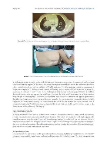
Figure 1. CT scan showing diffuse consolidation and bronchiectasis of the right upper lobe due to severe bronchomalacia
Figure 2. Left decubitus position with the right hand fixed to the right ear to open the axillary space
in its beginning and is rarely performed. The surgical literature contains very few cases, which have been
conducted only by experts in this field, and most centers in the world still adopt the traditional methods,
[1,2]
either open thoracotomy or the multiportal VATS technique . After gaining extensive experience in
single-port surgery in all its forms in adults and performing it at an advanced level, we started to apply this
[3-7]
technique in children as well . One of the challenges a surgeon may face during a lobectomy in children
through the intercostal approach is the small space between the ribs, which may make the instrumentation
[7]
very difficult and challenging . Therefore, we found that it makes sense to perform this type of operation via
the subxiphoid approach, which may provide more space for the instrumentation in addition to comfortable
angles for the instruments during the dissection of the hilum. In this article, we report the first case of
subxiphoid uniportal VATS lobectomy conducted for a 2.5-year-old child, and we review some of the
observations we found during the surgery.
CASE PRESENTATION
A thirty-month-old male patient suffered from recurrent chest infections since birth, which necessitated
several hospital admissions and antibiotics therapy. The chest CT scan showed right upper lobe
consolidation and bronchiectasis [Figure 1]. Bronchoscopy was performed to rule out any intrinsic factor or
other associated anomalies. The procedure showed a significant narrowing of the right upper lobe bronchus
due to severe bronchomalacia. The echocardiogram showed no cardiac abnormalities. The multidisciplinary
team forum decided that lobectomy is indicated.
Surgical technique
The operation was performed under general anesthesia. Isolated right lung ventilation was obtained by
advancing an uncuffed single-lumen endotracheal tube to the left main bronchus. The baby was positioned