
Page 38 - Read Online
P. 38
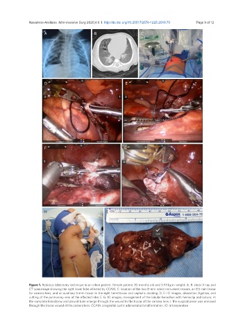
Navarrete-Arellano. Mini-invasive Surg 2020;4:9  I  http://dx.doi.org/10.20517/2574-1225.2019.70                             Page 9 of 12

                       A                       B                      C


                       D                                      E


                       F                                     G


                       H                             I


               Figure 1. Robotics lobectomy technique in an infant patient. Female patient, 10 months old and 5.93 kg in weight. A, B: chest X-ray and
               CT scan image showing the right lower lobe affected by CCAM; C: location of the two 8-mm robot instrument trocars, an 8.5-mm trocar
               for camera lens, and an auxiliary 5-mm trocar in the right hemithorax and cephalic docking; D, E: IO images, dissection, ligation, and
               cutting of the pulmonary vein of the affected lobe; F, G: IO images, management of the lobular bronchus with hemoclip and suture; H:
               the complete lobectomy and pleural tube emerge through the wound to the trocar of the camera lens; I: the surgical piece was removed
               through the trocar wound of the camera lens. CCAM: congenital cystic adenomatoid malformation; IO: Intraoperative