
Page 158 - Read Online
P. 158
Sollie et al. Mini-invasive Surg 2020;4:80 I http://dx.doi.org/10.20517/2574-1225.2020.81 Page 3 of 9
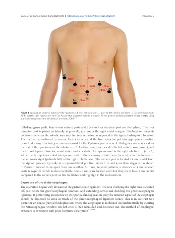
Figure 1. Location of ports for robotic Heller myotomy. LR: liver retractor port; L: port for left robotic arm (arm 1); C: camera port (arm
2); R: port for right robotic arm (arm 3); Ac: port for accessory robotic arm (arm 4); As: port for bedside assistant. Image created using
public domain photo from Wikipedia Commons, 2008 [23]
rolled-up gauze pads. Four 8-mm robotic ports and a 5-mm liver retractor port are then placed. The liver
retractor port is placed as laterally as possible, just under the right costal margin. This location prevents
collisions between the robotic arm and the liver retractor, as opposed to the typical subxiphoid location.
The patient is positioned in reverse Trendelenberg and the liver retractor put into appropriate position
prior to docking. The 0-degree camera is used for the Optiview port access. A 30-degree camera is used for
the rest of the operation via the robotic arm 2. Cadiere forceps are used in the left robotic arm (arm 1), and
the curved bipolar dissector, vessel sealer, and fenestrated forceps are used in the right robotic arm (arm 3),
while the tip-up fenestrated forceps are used in the accessory robotic arm (arm 4), which is located to
the surgeon’s right (patient’s left) of the right robotic arm. The camera port is located 15 cm caudal from
the xiphoid process, typically in a supraumbilical position. Arms 1, 3, and 4 are then staggered as shown
in Figure 1, located 9 cm apart from one another. At times, in small patients, a distance of 8 cm between
ports is required which is also acceptable. Arms 1 and 3 are located such that they are at least 3 cm cranial
compared to the camera port, as this facilitates working high in the mediastinum.
Exposure of the distal esophagus
The operation begins with division of the gastrohepatic ligament. The area overlying the right crus is cleared
off, just below the gastroesophageal junction, and extending across and dividing the phrenoesopahgeal
ligament. If performing an anterior or Dor partial fundoplication, only the anterior aspect of the esophagus
should be dissected to leave as much of the phrenoesophageal ligament intact. This is in contrast to a
posterior or Toupet partial fundoplication where the esophagus is mobilized circumferentially by clearing
the retroesophageal window. The left crus is then identified and dissected out. The method of esophageal
exposure is consistent with prior literature description [17,24-26] .