
Page 146 - Read Online
P. 146
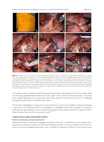
Khaitan et al. Mini-invasive Surg 2020;4:51  I  http://dx.doi.org/10.20517/2574-1225.2020.34                                       Page 5 of 14


               Figure 2. Thoracic portion of robotic Ivor Lewis esophagectomy. Robotic ports are placed in the right chest as shown [A: (c) camera
               port; (a) assistant port]; dissection in the chest typically begins with division of the inferior pulmonary ligament (B) followed by
               circumferential dissection of the esophagus (C) to allow placement of a Penrose drain around it (D); if performing the operation for an
               esophageal malignancy, nodal tissue is swept up with the specimen; the airway will be visualized during the dissection (BI). The azygos
               vein is divided flush with the cava (E) and the esophagus is transected superior to the azygos (F); the gastric conduit is delivered into
               the chest (G); and the proximal esophageal margin is checked in malignant cases (H); a transoral anvil is then delivered through the
               esophageal staple line (I) and an end-to-end anastomotic stapler used to complete the anastomosis, performed extra-corporeally here
               (images not captured). BI: bronchus intermedius

               A nasogastric tube is carefully advanced beyond the anastomosis and the gastrotomy site is resected using
               an endoscopic gastrointestinal anastomosis (GIA) stapler. After creation of the anastomosis, a soft tissue
               drain is placed adjacent to the conduit, and a chest tube is placed in the pleural cavity, prior to re-inflating
               the right lung under direct visualization and closure.


               Of note, the esophagogastric anastomosis can be performed in any of several different manners, including
               a linear side-to-side (functional end-to-end) stapled or a completely sewn 2-layer technique. No particular
               method has proven superior in terms of anastomotic leakage, though stapled anastomoses appear to lead to
               fewer strictures than ones that are completely sewn [19,20] .

               THREE-FIELD RAMIE (MCKEOWN RAMIE)
               Patient positioning and port placement
               McKeown RAMIE is started with esophageal dissection in the chest. A double lumen endotracheal tube is
               placed and the patient is rotated to the left lateral decubitus position. The lung is isolated and four robotic
               ports are used for esophageal mobilization with or without an assistant port, similar to the thoracic phase