
Page 144 - Read Online
P. 144
Khaitan et al. Mini-invasive Surg 2020;4:51 I http://dx.doi.org/10.20517/2574-1225.2020.34 Page 3 of 14
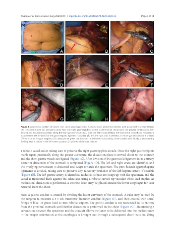
Figure 1. Abdominal portion of robotic Ivor Lewis esophagectomy. A depiction of abdominal robotic port placement is demonstrated
[A: (c) camera port; (a) assistant port]; first, the right gastroepiploic arcade is defined (B, red arrow); the greater omentum is then
divided and dissection proceeds along the short gastric vessels (C); once the left crus is defined, the stomach is rotated and retrogastric
attachments are divided (D); the gastrohepatic ligament is divided (E) and the right crus is defined; a 4-5 cm gastric conduit is created
(F) with serial firing of staplers (G); indocyanine green can be used to define the vascularity of the conduit (H); finally, a jejunostomy
feeding tube is placed in the left lower quadrant (I) prior to abdominal closure
a robotic vessel sealer, taking care to preserve the right gastroepiploic arcade. Once the right gastroepiploic
trunk tapers proximally along the greater curvature, the dissection plane is moved closer to the stomach
and the short gastric vessels are ligated [Figure 1C]. After division of the gastrocolic ligament in its entirety,
posterior dissection of the stomach is completed [Figure 1D]. The left and right crura are identified and
the overlying peritoneum is dissected and swept towards the specimen. The pars flaccida (gastrohepatic
ligament) is divided, taking care to preserve any accessory branches of the left hepatic artery, if sizeable
[Figure 1E]. The left gastric artery is identified, nodes at its base are swept up with the specimen, and the
vessel is transected flush against the celiac axis using a robotic curved tip vascular white load stapler. As
mediastinal dissection is performed, a Penrose drain may be placed around the lower esophagus for later
retrieval from the chest.
Next, a gastric conduit is created by dividing the lesser curvature of the stomach. A ruler may be used by
the surgeon to measure a 4-5 cm transverse diameter conduit [Figure 1F], and then created with serial
firings of blue- or green-load 45 mm robotic staplers. The gastric conduit is not transected in its entirety
from the proximal stomach until further dissection is performed in the chest [Figure 1G]. Maintaining a
connection between the specimen and the conduit allows the latter to be delivered into the mediastinum
in the proper orientation as the esophagus is brought out through a subsequent chest incision. Using