
Page 90 - Read Online
P. 90
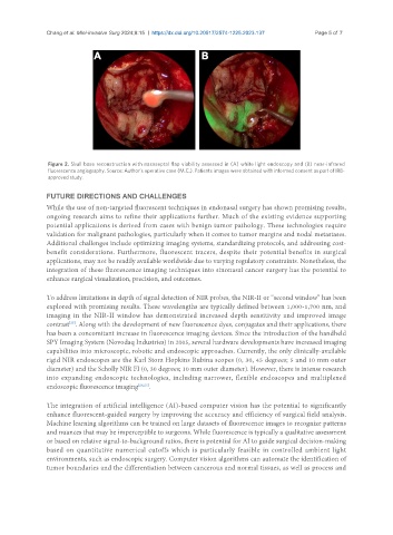
Chang et al. Mini-invasive Surg 2024;8:15  https://dx.doi.org/10.20517/2574-1225.2023.137  Page 5 of 7


                Figure 2. Skull base reconstruction with nasoseptal flap viability assessed in (A) white light endoscopy and (B) near-infrared
                fluorescence angiography. Source: Author’s operative case (M.C.). Patients images were obtained with informed consent as part of IRB-
                approved study.

               FUTURE DIRECTIONS AND CHALLENGES
               While the use of non-targeted fluorescent techniques in endonasal surgery has shown promising results,
               ongoing research aims to refine their applications further. Much of the existing evidence supporting
               potential applications is derived from cases with benign tumor pathology. These technologies require
               validation for malignant pathologies, particularly when it comes to tumor margins and nodal metastases.
               Additional challenges include optimizing imaging systems, standardizing protocols, and addressing cost-
               benefit considerations. Furthermore, fluorescent tracers, despite their potential benefits in surgical
               applications, may not be readily available worldwide due to varying regulatory constraints. Nonetheless, the
               integration of these fluorescence imaging techniques into sinonasal cancer surgery has the potential to
               enhance surgical visualization, precision, and outcomes.

               To address limitations in depth of signal detection of NIR probes, the NIR-II or “second window” has been
               explored with promising results. These wavelengths are typically defined between 1,000-1,700 nm, and
               imaging in the NIR-II window has demonstrated increased depth sensitivity and improved image
               contrast . Along with the development of new fluorescence dyes, conjugates and their applications, there
                      [25]
               has been a concomitant increase in fluorescence imaging devices. Since the introduction of the handheld
               SPY Imaging System (Novodaq Industries) in 2005, several hardware developments have increased imaging
               capabilities into microscopic, robotic and endoscopic approaches. Currently, the only clinically-available
               rigid NIR endoscopes are the Karl Storz Hopkins Rubina scopes (0, 30, 45 degrees; 5 and 10 mm outer
               diameter) and the Scholly NIR FI (0, 30 degrees; 10 mm outer diameter). However, there is intense research
               into expanding endoscopic technologies, including narrower, flexible endoscopes and multiplexed
               endoscopic fluorescence imaging [26,27] .


               The integration of artificial intelligence (AI)-based computer vision has the potential to significantly
               enhance fluorescent-guided surgery by improving the accuracy and efficiency of surgical field analysis.
               Machine learning algorithms can be trained on large datasets of fluorescence images to recognize patterns
               and nuances that may be imperceptible to surgeons. While fluorescence is typically a qualitative assessment
               or based on relative signal-to-background ratios, there is potential for AI to guide surgical decision-making
               based on quantitative numerical cutoffs which is particularly feasible in controlled ambient light
               environments, such as endoscopic surgery. Computer vision algorithms can automate the identification of
               tumor boundaries and the differentiation between cancerous and normal tissues, as well as process and