
Page 89 - Read Online
P. 89
Page 4 of 7 Chang et al. Mini-invasive Surg 2024;8:15 https://dx.doi.org/10.20517/2574-1225.2023.137
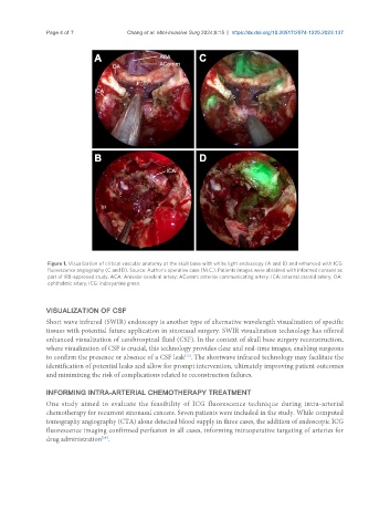
Figure 1. Visualization of critical vascular anatomy at the skull base with white light endoscopy (A and B) and enhanced with ICG
fluorescence angiography (C and D). Source: Author’s operative case (M.C.). Patients images were obtained with informed consent as
part of IRB-approved study. ACA: Anterior cerebral artery; AComm: anterior communicating artery; ICA: internal carotid artery; OA:
ophthalmic artery; ICG: indocyanine green.
VISUALIZATION OF CSF
Short wave infrared (SWIR) endoscopy is another type of alternative wavelength visualization of specific
tissues with potential future application in sinonasal surgery. SWIR visualization technology has offered
enhanced visualization of cerebrospinal fluid (CSF). In the context of skull base surgery reconstruction,
where visualization of CSF is crucial, this technology provides clear and real-time images, enabling surgeons
[23]
to confirm the presence or absence of a CSF leak . The shortwave infrared technology may facilitate the
identification of potential leaks and allow for prompt intervention, ultimately improving patient outcomes
and minimizing the risk of complications related to reconstruction failures.
INFORMING INTRA-ARTERIAL CHEMOTHERAPY TREATMENT
One study aimed to evaluate the feasibility of ICG fluorescence technique during intra-arterial
chemotherapy for recurrent sinonasal cancers. Seven patients were included in the study. While computed
tomography angiography (CTA) alone detected blood supply in three cases, the addition of endoscopic ICG
fluorescence imaging confirmed perfusion in all cases, informing intraoperative targeting of arteries for
drug administration .
[24]