
Page 23 - Read Online
P. 23
Page 6 of 8 Hashida et al. Mini-invasive Surg 2024;8:14 https://dx.doi.org/10.20517/2574-1225.2023.139
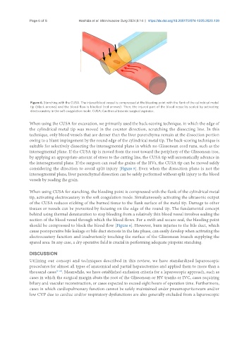
Figure 6. Stanching with the CUSA. The injured blood vessel is compressed at the bleeding point with the flank of the cylindrical metal
tip (black arrows) and the blood flow is blocked (red arrows). Then, the injured part of the blood vessel is sealed by activating
electrocautery in the soft coagulation mode. CUSA: Cavitron ultrasonic surgical aspirator.
When using the CUSA for excavation, we primarily used the back-scoring technique, in which the edge of
the cylindrical metal tip was moved in the counter direction, scratching the dissecting line. In this
technique, only blood vessels that are denser than the liver parenchyma remain at the dissection portion
owing to a blunt impingement by the round edge of the cylindrical metal tip. The back-scoring technique is
suitable for selectively dissecting the intersegmental plane in which no Glissonean cord runs, such as the
intersegmental plane. If the CUSA tip is moved from the root toward the periphery of the Glissonean tree,
by applying an appropriate amount of stress to the cutting line, the CUSA tip will automatically advance in
the intersegmental plane. If the surgeon can read the grains of the HVs, the CUSA tip can be moved safely
considering the direction to avoid split injury [Figure 5]. Even when the dissection plane is not the
intersegmental plane, liver parenchymal dissection can be safely performed without split injury to the blood
vessels by reading the grain.
When using CUSA for stanching, the bleeding point is compressed with the flank of the cylindrical metal
tip, activating electrocautery in the soft coagulation mode. Simultaneously activating the ultrasonic output
of the CUSA reduces sticking of the burned tissue to the flank surface of the metal tip. Damage to other
tissues or vessels can be prevented by focusing on the edge of the round tip. The fundamental concept
behind using thermal denaturation to stop bleeding from a relatively thin blood vessel involves sealing the
section of the blood vessel through which the blood flows. For a swift and secure seal, the bleeding point
should be compressed to block the blood flow [Figure 6]. However, burn injuries to the bile duct, which
cause postoperative bile leakage or bile duct stenosis in the late phase, can easily develop when activating the
electrocautery function and inadvertently touching the surface of the Glissonean branch supplying the
spared area. In any case, a dry operative field is crucial in performing adequate pinpoint stanching.
DISCUSSION
Utilizing our concept and techniques described in this review, we have standardized laparoscopic
procedures for almost all types of anatomical and partial hepatectomies and applied them to more than a
thousand cases [9-18] . Meanwhile, we have established exclusion criteria for a laparoscopic approach, such as
cases in which the surgical margin abuts the root of the Glissonean or HV trunks or IVC, cases requiring
biliary and vascular reconstruction, or cases expected to exceed eight hours of operation time. Furthermore,
cases in which cardiopulmonary function cannot be safely maintained under pneumoperitoneum and/or
low CVP due to cardiac and/or respiratory dysfunctions are also generally excluded from a laparoscopic