
Page 21 - Read Online
P. 21
Page 4 of 8 Hashida et al. Mini-invasive Surg 2024;8:14 https://dx.doi.org/10.20517/2574-1225.2023.139
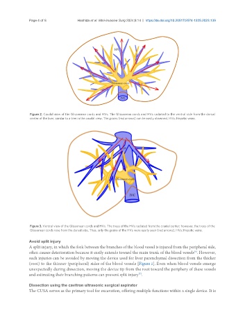
Figure 2. Caudal view of the Glissonean cords and HVs. The Glissonean cords and HVs radiated to the ventral side from the dorsal
center of the liver, similar to a tree in the caudal view. The grains (red arrows) can be easily observed. HVs: Hepatic veins.
Figure 3. Ventral view of the Glissonean cords and HVs. The trees of the HVs radiated from the cranial center; however, the trees of the
Glissonean cords rose from the dorsal side. Thus, only the grains of the HVs were easily seen (red arrows). HVs: Hepatic veins.
Avoid split injury
A split injury, in which the fork between the branches of the blood vessel is injured from the peripheral side,
often causes deterioration because it easily extends toward the main trunk of the blood vessels . However,
[8]
such injuries can be avoided by moving the device used for liver parenchymal dissection from the thicker
(root) to the thinner (peripheral) sides of the blood vessels [Figure 5]. Even when blood vessels emerge
unexpectedly during dissection, moving the device tip from the root toward the periphery of these vessels
[7]
and estimating their branching patterns can prevent split injury .
Dissection using the cavitron ultrasonic surgical aspirator
The CUSA serves as the primary tool for excavation, offering multiple functions within a single device. It is