
Page 45 - Read Online
P. 45
George et al. Mini-invasive Surg 2024;8:4 https://dx.doi.org/10.20517/2574-1225.2023.102 Page 11 of 23
positive rate
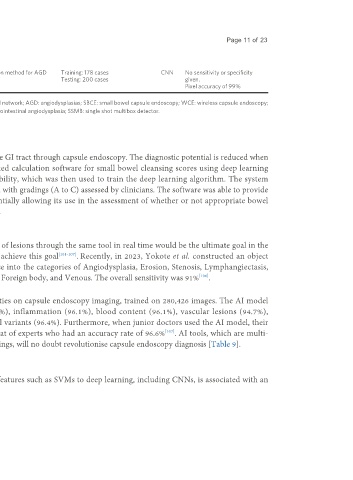
Chu Vascular lesions and 2023 Retrospective China Utilise CNN segmentation method for AGD Training: 178 cases CNN No sensitivity or specificity
[53]
et al. angiodysplasias detection Testing: 200 cases given.
Pixel accuracy of 99%
AI: Artificial intelligence; CE: capsule endoscopy; IPS: image-processed software; CNN: convolutional neural network; AGD: angiodysplasias; SBCE: small bowel capsule endoscopy; WCE: wireless capsule endoscopy;
KID: koulaouzidis-iakovidis database; MLP: multilayer perceptron; SVM: support vector machine; GIA: gastrointestinal angiodysplasia; SSMB: single shot multibox detector.
Other applications of AI in capsule endoscopy.
Automated calculation of bowel preparation quality
Effective and thorough bowel cleansing is essential for high quality images of the GI tract through capsule endoscopy. The diagnostic potential is reduced when
bowel preparation is inadequately performed. Nam et al. created an automated calculation software for small bowel cleansing scores using deep learning
algorithms. A five-step scoring system was developed based on mucosal visibility, which was then used to train the deep learning algorithm. The system
assigned an average cleansing score (ranging from 1 to 5), which was compared with gradings (A to C) assessed by clinicians. The software was able to provide
objective, automated cleansing scores for small bowel preparation, thus potentially allowing its use in the assessment of whether or not appropriate bowel
preparation has been achieved for small bowel pathology detection [Table 8].
[100]
Multiple lesion characterisation
A functioning, highly accurate method to detect and characterise a wide range of lesions through the same tool in real time would be the ultimate goal in the
foreseeable future for AI research. Various models have so far attempted to achieve this goal [101-107] . Recently, in 2023, Yokote et al. constructed an object
detection AI model from a dataset of 18,481 images to detect and characterise into the categories of Angiodysplasia, Erosion, Stenosis, Lymphangiectasis,
[106]
Lymph follicle, Submucosal tumour, Polyp‐like, Bleeding, Diverticula, Redness, Foreign body, and Venous. The overall sensitivity was 91% .
Also, in 2023, Ding et al. developed an AI model to detect various abnormalities on capsule endoscopy imaging, trained on 280,426 images. The AI model
showed high sensitivity in detecting various abnormalities: red spots (97.8%), inflammation (96.1%), blood content (96.1%), vascular lesions (94.7%),
protruding lesions (95.6%), parasites (100%), diverticulum (100%), and normal variants (96.4%). Furthermore, when junior doctors used the AI model, their
[107]
overall accuracy increased from 85.5% to 97.9% and became comparable to that of experts who had an accuracy rate of 96.6% . AI tools, which are multi-
faceted and have the ability to detect and characterise a variety of common findings, will no doubt revolutionise capsule endoscopy diagnosis [Table 9].
DISCUSSION
The shifting of utilised AI types over time from traditional machine learning features such as SVMs to deep learning, including CNNs, is associated with an
increase in accuracy, sensitivity and specificity of diagnostic results.