
Page 84 - Read Online
P. 84
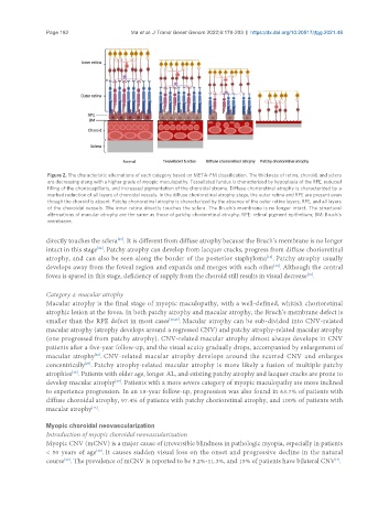
Page 182                  Ma et al. J Transl Genet Genom 2022;6:179-203  https://dx.doi.org/10.20517/jtgg.2021.48


                Figure 2. The characteristic alternations of each category based on META-PM classification. The thickness of retina, choroid, and sclera
                are decreasing along with a higher grade of myopic maculopathy. Tessellated fundus is characterized by hypoplasia of the RPE, reduced
                filling of the choriocapillaris, and increased pigmentation of the choroidal stroma. Diffuse chorioretinal atrophy is characterized by a
                marked reduction of all layers of choroidal vessels. In the diffuse chorioretinal atrophy stage, the outer retina and RPE are present even
                though the choroid is absent. Patchy chorioretinal atrophy is characterized by the absence of the outer retina layers, RPE, and all layers
                of the choroidal vessels. The inner retina directly touches the sclera. The Bruch’s membrane is no longer intact. The structural
                alternations of macular atrophy are the same as those of patchy chorioretinal atrophy. RPE: retinal pigment epithelium; BM: Bruch’s
                membrane.

               directly touches the sclera . It is different from diffuse atrophy because the Bruch’s membrane is no longer
                                     [23]
               intact in this stage . Patchy atrophy can develop from lacquer cracks, progress from diffuse chorioretinal
                               [24]
               atrophy, and can also be seen along the border of the posterior staphyloma . Patchy atrophy usually
                                                                                   [19]
               develops away from the foveal region and expands and merges with each other . Although the central
                                                                                     [19]
               fovea is spared in this stage, deficiency of supply from the choroid still results in visual decrease .
                                                                                               [19]
               Category 4: macular atrophy
               Macular atrophy is the final stage of myopic maculopathy, with a well-defined, whitish chorioretinal
               atrophic lesion at the fovea. In both patchy atrophy and macular atrophy, the Bruch’s membrane defect is
               smaller than the RPE defect in most cases [23,25] . Macular atrophy can be sub-divided into CNV-related
               macular atrophy (atrophy develops around a regressed CNV) and patchy atrophy-related macular atrophy
               (one progressed from patchy atrophy). CNV-related macular atrophy almost always develops in CNV
               patients after a five-year follow-up, and the visual acuity gradually drops, accompanied by enlargement of
               macular atrophy . CNV-related macular atrophy develops around the scarred CNV and enlarges
                              [26]
               concentrically . Patchy atrophy-related macular atrophy is more likely a fusion of multiple patchy
                           [27]
               atrophies . Patients with older age, longer AL, and existing patchy atrophy and lacquer cracks are prone to
                       [15]
               develop macular atrophy . Patients with a more severe category of myopic maculopathy are more inclined
                                    [19]
               to experience progression. In an 18-year follow-up, progression was also found in 63.7% of patients with
               diffuse choroidal atrophy, 97.4% of patients with patchy chorioretinal atrophy, and 100% of patients with
                             [15]
               macular atrophy .

               Myopic choroidal neovascularization
               Introduction of myopic choroidal neovascularization
               Myopic CNV (mCNV) is a major cause of irreversible blindness in pathologic myopia, especially in patients
               < 50 years of age . It causes sudden visual loss on the onset and progressive decline in the natural
                              [28]
               course . The prevalence of mCNV is reported to be 5.2%-11.3%, and 15% of patients have bilateral CNV .
                                                                                                        [4]
                     [26]