
Page 88 - Read Online
P. 88
Page 186 Ma et al. J Transl Genet Genom 2022;6:179-203 https://dx.doi.org/10.20517/jtgg.2021.48
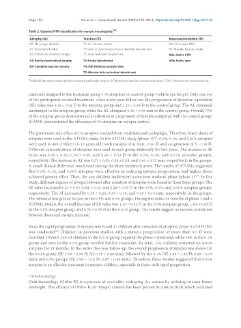
Table 2. Updated ATN classification for myopic maculopathy [80]
Atrophy (A) Traction (T) Neovascularization (N)
A0: No myopic atrophy T0: No macular schisis N0: No myopic CNV
A1: Tessellated fundus T1: Inner or outer foveoschisis or lamellar macular hole N1: Macular lacquer cracks
A2: Diffuse chorioretinal atrophy T2: Inner and outer foveoschisis N2a: Active CNV
A3: Patchy chorioretinal atrophy T3: Foveal detachment N2b: Fuchs’ spot
A4: Complete macular atrophy T4: Full-thickness macular hole
T5: Macular hole and retinal detachment
The bold fonts mark stages defined as severe pathologic myopia. ATN: atrophy-traction-neovascularization. CNV: choroidal neovascularization.
randomly assigned to the treatment group (1% atropine) or control group (vehicle eye drops). Only one eye
of the participants received treatment. After a two-year follow-up, the progression of spherical equivalent
(SE) value was 0.28 ± 0.92 D in the atropine group and 1.20 ± 0.69 D in the control group. The AL remained
unchanged in the atropine group, while the AL elongated 0.38 ± 0.38 mm in the control group. Overall, 77%
of the atropine group demonstrated a reduction in progression of myopia compared with the control group.
ATOM1 demonstrated the efficiency of 1% atropine on myopia control.
The prominent side effect of 1% atropine resulted from mydriasis and cycloplegia. Therefore, lower doses of
atropine were used in the ATOM2 study. In the ATOM2 study (phase 1) , 0.5%, 0.1%, and 0.01% atropine
[82]
were used in 400 children (6-12 years old) with myopia of at least -2.00 D and astigmatism of ≤ -1.50 D.
Different concentrations of atropine were used in each group bilaterally for two years. The increase in SE
value was 0.30 ± 0.60, 0.38 ± 0.60, and 0.49 ± 0.63 D in the 0.5%, 0.1%, and 0.01% atropine groups,
respectively. The increase in AL was 0.27 ± 0.25, 0.28 ± 0.28, and 0.41 ± 0.32 mm, respectively, in the groups.
A small clinical difference was found among the three treatment arms. The results of ATOM2 suggested
that 0.5%, 0.1%, and 0.01% atropine were effective in reducing myopia progression, and higher doses
achieved greater effect. Then, the 400 children underwent a one-year washout phase (phase 2) . In this
[83]
study, different degrees of myopic rebound after cessation of atropine were found in these three groups. The
SE value increased 0.87 ± 0.52, 0.68 ± 0.45, and 0.28 ± 0.33 D in the 0.5%, 0.1%, and 0.01% atropine groups,
respectively. The Al increased by 0.35 ± 0.20, 0.33 ± 0.18, and 0.19 ± 0.13 mm, respectively, in the groups.
The rebound was greater in eyes in the 0.5% and 0.1% groups. During the entire 36 months of phase 1 and 2
ATOM2 studies, the overall increase of SE value was 1.15 ± 0.81 D in the 0.5% atropine group, 1.04 ± 0.83 D
in the 0.1% atropine group, and 0.72 ± 0.72 D in the 0.01% group. The results suggest an inverse correlation
between doses and myopic increase.
Since the rapid progression of myopia was found in children after cessation of atropine, phase 3 of ATOM2
was conducted . Children in previous studies with a myopic progression of more than 0.5 D were
[84]
recruited. Overall, 24% of children in the 0.01% group required the phase 3 treatment, while 59% in the 0.1%
group and 68% in the 0.5% group needed further treatment. In total, 192 children restarted on 0.01%
atropine for 24 months. In the entire five-year follow-up, the overall progression of myopia was slowest in
the 0.01% group (SE 1.38 ± 0.98 D; AL 0.75 ± 0.48 mm), followed by the 0.1% (SE 1.83 ± 1.16 D; 0.85 ± 0.53
mm) and 0.5% groups (SE 1.98 ± 1.10 D; 0.87 ± 0.49 mm). Therefore, these studies suggested that 0.01%
atropine is an effective treatment in myopic children, especially in those with rapid progression.
Orthokeratology
Orthokeratology (Ortho-K) is a process of reversibly reshaping the cornea by utilizing contact lenses
overnight. The efficacy of Ortho-K on myopic control has been proved in clinical trials which recruited