Page 104 - Read Online
P. 104

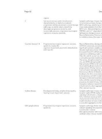
Page 92                                                                               Dewsbury et al. J Transl Genet Genom 2024;8:85-101      https://dx.doi.org/10.20517/jtgg.2023.58


                                                    regions
                              C                     Sub-acute nervous system involvement,     Synaptic pathology (mouse model); impairment of SNARE function; mitochondrial cholesterol accumulation; the   [59]
                                                    characterized by a relatively moderate    simultaneous primary storage of cholesterol, coupled with the secondary storage of sphingomyelin, is a prime driver for   [60]
                                                    progression and less prominent visceral storage  NPC1 pathology, interfering with TRPML1 and TRPML1-dependent maintenance of lysosomal homeostasis. Sphingolipids   [61]
                                                    pathology; cerebellar ataxia, dysarthria,   show mislocalization from the Golgi apparatus to lysosomes, as demonstrated by impaired trafficking of lactosylceramide in  [62]
                                                    dysphagia, progressive dementia, and      NPC1 cells. Abnormal lipid accumulation in NPC1 patient lysosomes results in secondary lysosomal storage by blocking   [63]
                                                                                                            2+
                                                    occasionally seizures; progressive neurological   TRPML1- and Ca -dependent lysosomal trafficking; this storage could be reverted by the TRPML agonist ML-SA1.   [61]
                                                                                                                                                                                     +    2+
                                                    regression, seizures, spasticity          Sphingosine storage induces calcium depletion in lysosomes, possibly through an inhibitory effect on Na /Ca  exchangers.  [63]
                                                                                              Neuroinflammation; Peroxisomal dysfunction; Decreased oxidative respiration/reduced ATP levels; Increased vulnerability   [64]
                                                                                              to oxidative stress; Decrease in mitochondrial GSH resulting in Cytochrome c release                      [65,66]
                                                                                                                                                                                                        [67]
                                                                                                                                                                                                        [68]
                              Gaucher disease I-III  Progressive neurological regression, seizures,   Neuroinflammation, dysregulated calcium homeostasis, decreased mitochondrial membrane potential, selective reduction of  [64]
                                                    spasticity (mainly III),                  OXPHOS complexes, accumulation of APP and α-synuclein, Reduced O  consumption/reduced ATP levels          [65,66]
                                                                                                                                                          2
                                                    cognitive impairment, psychiatric disturbances  Significant systemic oxidative stress demonstrated by altered GSH status and lowered catalase enzymatic activity, as well as  [58]
                                                    (GD I and III)                            elevated lipid peroxidation (GD I) (human)                                                                [69]
                                                                                              Increased Tmem119 mRNA expression leads to abnormal microglia growth and proliferation, as well as increased Iba1   [70]
                                                                                              expression exhibiting a higher degree of microglial cell activation; correlated by increases in pro-inflammatory cytokines   [71]
                                                                                              Tnfa, Il1b, and Il6, as well as chemokines Cxcl10, Ccl2, Ccl5, and Cxcl9. Markedly raised antigen-presenting cell receptor Cd86,  [72]
                                                                                              a cell surface protein identifying M1 microglia (adult genetic nGD model)                                 [73]
                                                                                              The active, phosphorylated form of c-Abl is increased, and interacts with RIPK3, which is in turn phosphorylated at a tyrosine  [74]
                                                                                              site, and RIPK3 phosphorylation is reduced when c-Abl is inhibited. This shows that c-Abl signaling is the upstream pathway  [75]
                                                                                              that activates RIPK3 in GD (adult human and mice GD models)                                               [76]
                                                                                              Accumulating Β-glucosylceramide in GD activated microglia through macrophage-inducible C-type lectin induces   [77]
                                                                                              phagocytosis of living neurons, exacerbating symptoms of GD. This is augmented by tumor necrosis factor (TNF) secreted   [78]
                                                                                              from activated microglia that sensitizes neurons for phagocytosis (human model)
                                                                                              Deleterious effects of Wnt/β-catenin downregulation in neuronopathic GD may be ameliorated by the prevention of Dkk1
                                                                                              binding to the Wnt co-receptor LRP6 (mouse model)
                                                                                              In the presence of GBA1 mutations, increased chaperone and LONP1 activity may promote the degradation of
                                                                                              damaged/misfolded intramitochondrial GCase. Complex I activity is ameliorated with LONP1 inhibition in GBA1 mutant HEK
                                                                                              cells, with the opposite being observed in WT-Gcase cells. This is also observed in iPSC neurons, showing that in the
                                                                                              presence of mutant GCase, LONP1 proteolytic activity is more pronounced than its chaperone function. This increased
                                                                                              interaction between mutant GCase and LONP1 may also interfere with the folding properties of LONP1, leading to
                                                                                              mitochondrial protein aggregation, which may result in decreased CI activity and mitochondrial α-synuclein accumulation in
                                                                                              iPSC-derived neurons

                              Krabbe disease        Developmental delay, peripheral neuropathy,   Synaptic pathology (mouse model) Peroxisomal dysfunction - downregulates the peroxisome proliferator-activated   [79]
                                                                                                                                                                                                  2+
                                                    hearing/visual impairment, seizures       receptor-alpha (PPAR-alpha). Decreased mitochondrial membrane potential oxidative stress/GSH Dysregulation of Ca    [80,81]
                                                                                              signaling Cytochrome c release                                                                            [82]
                                                                                              GALC deletion causes growth and motor coordination defects, and inflammatory gliosis due to the significant accumulation  [83]
                                                                                              of psychosine in the nervous system. This was shown to result in profound neuro-axonal degeneration with a mild effect on   [84]
                                                                                              myelin structure (mouse model)                                                                            [85]
                                                                                              α-synuclein aggregation in the brain tissue to form fibrils (human)
                              GM1 gangliosidosis    Progressive neurological regression, seizures,   Synaptic pathology (feline model) Neuroinflammation; enhanced autophagy and mitochondrial dysfunction. Dysregulated   [86,87]
                                                    spasticity                                calcium signaling. Decreased mitochondrial membrane potential. Increased vulnerability to oxidative stress-induced   [88]
                                                                                              Cytochrome c release                                                                                      [89]
                                                                                              Microglial activation with increased levels of LC3 autophagy regulator; increased microglial activation and proliferation in the  [90]
                                                                                              cerebral cortex (mouse model)                                                                             [91]
   99   100   101   102   103   104   105   106   107   108   109