Page 102 - Read Online
P. 102

Dewsbury et al. J Transl Genet Genom 2024;8:85-101     https://dx.doi.org/10.20517/jtgg.2023.58                                                                              Page 91


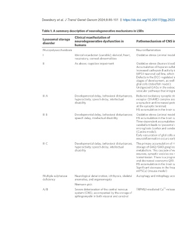
                              Table 1. A summary description of neurodegenerative mechanisms in LSDs

                                                    Clinical manifestation of
                              Lysosomal storage
                              disorder              neurodegenerative dysfunction in          Pathomechanism of CNS involvement                                                                         Reference
                                                    humans

                              Mucopolysaccharidosis                                           Neuroinflammation                                                                                         [33-35]
                              I                     Mental retardation (variable); skeletal, heart,   Oxidative stress (animal models and human blood)                                                  [36,37]
                                                    respiratory, corneal abnormalities
                              II                    As above; cognitive impairment            Oxidative stress (human blood)                                                                            [38]
                                                                                              Accumulation of heparan sulfate and dermatan sulfate in cerebrospinal fluid                               [39]
                                                                                              Increased cathepsin B activity in the brain tissue leads to leakage of this enzyme from the lysosome into the cytoplasm in a   [40]
                                                                                              MPSII neuronal cell line, which in turn activates the inflammasome pathway (mouse model);                 [41]
                                                                                              Defects in the DCC-regulated signaling pathway, lysosomal acidification, and Rab7 protein levels in neurons, affecting early  [42]
                                                                                              stages of development, as well as abnormalities in glial fibrillary acid protein (GFAP) levels, suggesting early activation of   [43]
                                                                                              glial cells (zebrafish model);
                                                                                              Undigested GAGs in the extracellular matrix lead to the impairment of the integrin involved in signaling axon guidance and
                                                                                              vesicular pathways that integrate neural circuit development in the early stages of neuronal development
                              III A                 Developmental delay, behavioral disturbances,   Reduced excitatory synaptic strength on the somatosensory cortex (mouse model); inhibition of soluble NSF attachment   [44]
                                                    hyperactivity; speech delay, intellectual   receptor (SNARE) complex assembly and synaptic vesicle recycling, possibly caused by perikaryal accumulation of insoluble  [45]
                                                    disability                                α-synuclein and increased proteasomal degradation of cysteine string protein α, resulting in low availability of these proteins  [46]
                                                                                              at the synaptic terminal;                                                                                 [47]
                                                                                              HS accumulation in the brain causes changes in oligodendrocyte cell state (zebrafish model)
                              III B                 Developmental delay, behavioral disturbances;   Oxidative stress (animal models); Golgi involvement;                                                [48,49]
                                                    speech delay, intellectual disability     HS accumulation in the brain causes changes in oligodendrocyte cell state (zebrafish model);              [46]
                                                                                              Time-dependent accumulation of HS and HS-NRE and progressive increase in LAMP1 staining in the forebrain and   [47]
                                                                                              cerebellum leads to lysosomal enlargement, atrophy of white matter, loss of Purkinje neurons, and progressive increase in   [50]
                                                                                              microgliosis (cortex and cerebellum) and astrogliosis (cerebellum). Raised Iba1 is an indicator of microglial activation   [51]
                                                                                              (Canine model);
                                                                                              Early vacuolation of glial cells and vacuolation of neurons, along with increased glial expression of GFAP and Iba1, suggest
                                                                                              neuroinflammation occurs early on in disease within the spinal cord and dorsal root ganglion (DRG) (Canine model)
                              III C                 Developmental delay, behavioral disturbances,   The primary accumulation of HS in microglial cells and neurons results in impaired autophagy, leading to secondary neuronal  [52]
                                                    hyperactivity; speech delay, intellectual   storage of GM2/GM3 gangliosides and misfolded proteins, neuroinflammation, and abnormalities in mitochondrial energy   [53]
                                                    disability                                metabolism. This cascade of events ultimately leads to neuronal death. Within the terminals of MPSIIIC hippocampal   [54]
                                                                                              neurons, synaptic vesicles are reduced; these neurons exhibit changes in distribution of excitatory synaptic markers and in   [55]
                                                                                              transmission. There is a progressive deficiency in mitochondrial function, with a selective reduction in OXPHOS complexes   [46]
                                                                                              and decreased coenzyme Q10.                                                                               [47]
                                                                                              HS accumulation in the brain causes changes in oligodendrocyte cell state (zebrafish model)               [54]
                                                                                              Significant decrease in the frequency and amplitude of excitatory and inhibitory miniature synaptic events (mEPSCs and
                                                                                              mIPSCs) (mouse model)
                              Multiple sulphatase   Neurological deterioration, ichthyosis, skeletal   Autophagy and mitophagy accumulation fragmentation decreased ATP content                         [55]
                              deficiency            anomalies, and organomegaly                                                                                                                         [56]
                                                    Niemann-pick
                                                                                                                2+
                              A/B                   Severe deterioration of the central nervous   TRPML1-mediated Ca -release is compromised                                                            [57]
                                                    system (CNS), accompanied by the storage of                                                                                                         [58]
                                                    sphingomyelin in both visceral and cerebral
   97   98   99   100   101   102   103   104   105   106   107