
Page 46 - Read Online
P. 46
Dasgupta et al. J Transl Genet Genom 2018;2:15. I https://doi.org/10.20517/jtgg.2018.21 Page 7 of 15
A B
C D
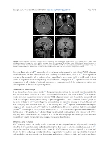
Figure 4. Typical magnetic resonance imaging features of group 4 medulloblastoma. Post-contrast axial T1-weighted (A) image show
a mildly enhancing tumor arising in the midline posterior fossa. Corresponding T2-weighted image (B) better delineates the extent of
primary tumor. Note the mild dilatation of the superior recess of the IVth ventricle on post-contrast sagittal T1-weighted (C) image and
presence of suprasellar/infundibular metastasis best appreciated on the post-contrast coronal T1-weighted (D) image
[24]
However, Lastowska et al. reported weak or minimal enhancement in 3 of 6 (50%) WNT-subgroup
[28]
medulloblastoma. In their cohort of adult WNT-pathway medulloblastoma, Zhao et al. found significant
contrast enhancement in all 17 patients, which was either heterogeneous (82%) or solid (18%). In their
[31]
cohort of 17 patients with WNT-pathway medulloblastoma, Dasgupta et al. reported intense contrast
enhancement in all patients; 53% showed homogeneous enhancement, while the enhancement pattern was
inhomogeneous in the remaining 47% of patients.
Intra-tumoral hemorrhage
[34]
It has been shown from animal studies that paracrine signals from the mutant β-catenin result in the
[34]
aberrant fenestrated vasculature in WNT-driven medulloblastoma. The same authors also reported
results from two independent cohorts wherein WNT-subgroup tumors were found to be associated with
frank hemorrhage in 90% of patients during surgery as opposed to 12.5% for the other three subgroups. In
[26]
the series by Patay et al. , hemorrhage was appreciated on pre-operative imaging in 31% of children with
[27]
WNT-subgroup medulloblastoma (n = 16). On the contrary, Keil et al. , reported absence of hemorrhage on
imaging in all 4 cases of adult WNT-pathway medulloblastoma. However, in another study involving adult
[28]
patients , hemorrhage or mineralization was reported in 11 of 17 (67%) WNT-pathway medulloblastoma.
[31]
In the series by Dasgupta et al. , hemorrhage was identified on imaging in 18% patients with WNT-
subgroup medulloblastoma (n = 17) compared to < 10% for other subgroups, necessitating the routine use of
susceptibility-weighted or gradient-echo imaging for reliable identification.
Other imaging features
WNT subgroup tumors are usually smaller in size and volume compared to other subgroups which may be
[27]
reflective of slow growth kinetics and indolent biology. In their series of adult medulloblastoma, Keil et al.
3
3
reported the median tumor volume to be 5.6 cm for WNT-subgroup tumors compared to 30.6 cm and
3
25 cm for SHH and group 4 medulloblastoma respectively. The authors also reported the absence of
hydrocephalus, macrometastasis, and hemorrhage as having very high specificity and positive predictive