
Page 44 - Read Online
P. 44
Dasgupta et al. J Transl Genet Genom 2018;2:15. I https://doi.org/10.20517/jtgg.2018.21 Page 5 of 15
Table 2. Typical imaging characteristics of individual molecular subgroups of medulloblastoma
MRI features WNT-MB SHH-MB Group 3 MB Group 4 MB
Location (horizontal Midline, but commonly extends to Lateralised location involving Midline location Midline location
axis) CP/CPA cistern hemispheres; midline location involving the IVth involving the IVth
common in infants/young children ventricle/vermis ventricle/vermis
Location (vertical Central in location, but can extend Superior location highly specific Central in location, but Inferior in location with
axis) inferiorly sometimes (often reaching or abutting the can extend inferiorly also dilatation of superior
tentorium) recess of IVth ventricle
Relation with dorsal Often seen infiltrating the dorsal Over 50% tumors away from Closely related and abuts Closely related and abuts
brainstem brainstem dorsal brainstem the dorsal brainstem the dorsal brainstem
Contrast- Homogeneous bright enhancement Variable pattern with moderate Heterogeneous “fluffy” Heterogeneous “patchy”
enhancement involving majority of the tumor enhancement type of enhancement type of enhancement
T2-weighted Mostly isointense and Mostly isointense and Mostly hypointense and Hyperintense/isointense
characteristics homogeneous heterogeneous homogeneous and homogeneous
Peri-tumoral edema Mild or absent Significant edema, often > 1.5 cm Absent or mild Absent or mild
beyond the tumor
Intra-tumoral Can be present Absent Absent Absent
hemorrhage
Cyst (size and Microcysts; intra-tumoral Microcysts & macrocysts; intra- Macrocyst; peri-tumoral Microcysts; intra-
location) tumoral, peri-tumoral tumoral
Hydrocephalus Generally absent (if present, mild Seldom seem (if present, mild to Moderate to severe Moderate to severe
to moderate) moderate) hydrocephalus hydrocephalus
Metastases Absent (rarely, if ever seen) Variable incidence Highest incidence Moderate incidence
(incidence, location, Spinal & posterior fossa Spinal metastases Suprasellar/infundibular
and pattern) Possible multi-centricity Laminar metastases Nodular metastases
MRI: magnetic resonance imaging; MB: medulloblastoma; WNT: wingless; SHH: sonic hedge hog; CP/CPA: cerebellar peduncle/
cerebellopontine angle
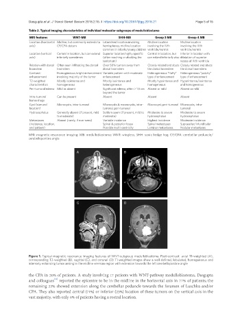
A B
C D
Figure 1. Typical magnetic resonance imaging features of WNT-subgroup medulloblastoma. Post-contrast axial T1-weighted (A),
corresponding T2-weighted (B), sagittal (C), and coronal (D) T1-weighted images show a well-defined, lobulated, homogeneous and
intensely enhancing tumor arising in the midline vermian region with extension towards the left cerebellopontine angle
the CPA in 20% of patients. A study involving 17 patients with WNT-pathway medulloblastoma, Dasgupta
and colleagues reported the epicentre to be in the midline in the horizontal axis in 77% of patients; the
[31]
remaining 23% showed extension along the cerebellar peduncle towards the foramen of Luschka and/or
CPA. They also reported central (71%) or inferior (23%) location of these tumors on the vertical axis in the
vast majority, with only 6% of patients having a rostral location.