
Page 43 - Read Online
P. 43
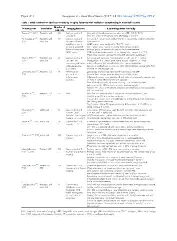
Page 4 of 15                                             Dasgupta et al. J Transl Genet Genom 2018;2:15. I  https://doi.org/10.20517/jtgg.2018.21

               Table 1. Brief summary of studies correlating imaging features with molecular subgrouping in medulloblastoma
               Author & year  Population  Number of   Imaging features   Key findings from the study
                                      patients
                     [21]
               Teo et al. , 2013  Pediatric MB  60  Conventional MRI   Hemispheric location was associated with SHH-MB (> 50%)
                                             (location)     Non-WNT/non-SHH tumors were lateralised in only 5%
               Perreault et al. [22] ,  Pediatric and   99   Conventional MRI   Group 3 & 4 tumors were predominantly located in the midline within the
               2014         adult MB         features; diffusion-  IVth ventricle
                                             weighted imaging   WNT tumors were localised to CP/CPA cistern
                                             including apparent   SHH tumors were having cerebellar hemispheric location
                                             diffusion coefficient   Midline group 4 tumors had no or minimal enhancement
                                             (ADC maps)     MRI based regression model correctly predicted subgroup in 65%
                                                            Mean ADC was not significantly different among the subgroups
               Wefers et al. [23] ,   Pediatric and   71  Conventional MRI   Subgroup with exclusive intracerebellar growth was SHH (52%)
               2014         adult MB         (location and   Most group 3 & 4 tumors grew in the midline in vermis (> 70%)
                                             relationship of tumor  A third of non-SHH tumors had tumor in caudal cerebellum
                                             with surrounding   Brainstem contact was seen in only 48% of SHH-MB as compared to 75%
                                             structures)    or more for other subgroups
               Lastowska et al. [24] ,  Pediatric MB  76  MRI (location   Lateralized location was significantly associated with SHH
               2015                          and contrast   None of SHH tumors were invading the brainstem floor
                                             enhancement    All group 3 tumors were associated with extensive contrast enhancement
                                             features)      (> 75% of tumor showing contrast uptake)
                                                            Over 60% of group 4 tumors were having no or minimal contrast
                                                            enhancement (< 10% of tumor showing contrast uptake)
                                                            For non-WNT/non-SHH tumors, extensive contrast uptake was associated
                                                            with poor survival
               Bluml et al. [25] ,   Pediatric MB  30  MRS  SHH-MB was associated with prominent choline/lipid peaks, low
               2016                                         creatinine, and little or no taurine levels.
                                                            Group 3 & 4 tumors were characterized by low lipd levels, high creatinine,
                                                            and readily detected taurine
                                                            The 5-metabolite MRS signature reliably differentiated SHH-MB from
                                                            group 3/group 4 tumors
               Patay et al. [26] ,   WNT MB  16  Conventional MRI   Involvement of foramen of Luschka, IVth ventricle, cisterna magna, and
               2015                          features (also   CPA was seen in WNT-MB
                                             assessed post-surgery  In 87% of patients, contrast enhancement involved entire tumor
                                             imaging for location) Intra-tumoral hemorrhage was seen in 31% of patients
               Keil et al. [27] , 2017 Adult MB  28  Conventional MRI   Absence of hydrocephalus, macrometastases, and hemorrohage was
                                             features       suggestive of WNT-MB
                                                            SHH-MB was associated with larger tumor volume and edema
                                                            Contact with lower rhombic lip and hemorrhage were relatively common
                                                            in group 4 tumors
               Zhao et al. [28] , 2017 Adult MB  125  Conventional MRI   Large majority of SHH-MB were hemispheric in location
                                             features       48% of SHH-MB had burden exclusively in rostral cerebellum
                                                            Peri-tumoral edema was seen commonly in SHH and WNT-MB
                                                            Group 4 tumors were predominant midline vermian in location
                                                            Minimal or no enhancement was seen in 50% of group 4 tumors
               Mata-Mbemba      Pediaric MB  119  Conventional MRI   Primary tumors in SHH-MB were hemispheric in location
               et al. [29] , 2018            features and diffusion  Primary tumors were smaller (< 3.5 cm) in group 3 MB compared to group
                                             imaging        4 and SHH-MB
                                                            Laminar metastates were commonly seen in group 3 tumors
                                                            Nodular metastases were commonly seen in group 4 tumors
                                                            Suprasellar/infundibular metastases were specific for group 4 MB
               Zapotocky et al. [30] ,  Metastatic   40  Conventional MRI   Cerebellar peripheral location was very common in SHH-MB
               2018         MB               features and diffusion  Minimal enhancement was seen in primary group 4 tumor
                                             imaging        CPA location was seen more commonly in WNT-MB
                                                            Spinal metastases were more commonly seen in group 3 tumors
                                                            Ependymal metastasis with restricted diffusion but no enhancement
                                                            (“mismatch pattern”) was seen in group 4 tumors (particualrly if located
                                                            in the infundibular recess)
               Dasgupta et al. ,  Pediatric and   111  Conventional   WNT-MB was asoociated with smaller tumor size, homogeneous contrast
                         [31]
               2018         adult MB         MRI features and   uptake, and intratumoral hemorrhage
                                             developed MRI-  SHH-MB was more likely to have lateralised and superior location, away
                                             based nomograms   from brainstem and presence of peri-tumoral edema
                                             for prediction of   Group 3 tumors were in midline location, with “fluffy” enhancement and
                                             subgroups      higher incidence of metatstatic diasease
                                                            Group 4 tumors were in midline and inferior in location with dilatation of
                                                            superior recess of IVth ventricle; showed minimal or “patchy” contrast
                                                            enhancement
               MRI: magnetic resonance imaging; MRS: magnetic resonance spectroscopy; MB: medulloblastoma; WNT: wingless; SHH: sonic hedge
               hog; ADC: apparent diffusion co-efficient; CP: cerebellar peduncle; CPA:cerebellopontine angle